Huimin Mao1, Weiqiang Dou2, Xinyi Wang1, Xinyu Wang1, Kunjian Chen1, and Yu Guo1
1The First Affiliated Hospital of Shandong First Medical University, Jinan, China, 2GE Healthcare, MR Research China, Beijing, P.R. China, Beijing, China
1The First Affiliated Hospital of Shandong First Medical University, Jinan, China, 2GE Healthcare, MR Research China, Beijing, P.R. China, Beijing, China
Intracranial vasculopathy is not a rare
complication after cranial irradiation, even in young patients. Patients after
cranial irradiation should be followed up with MR imaging including HR-MRI.
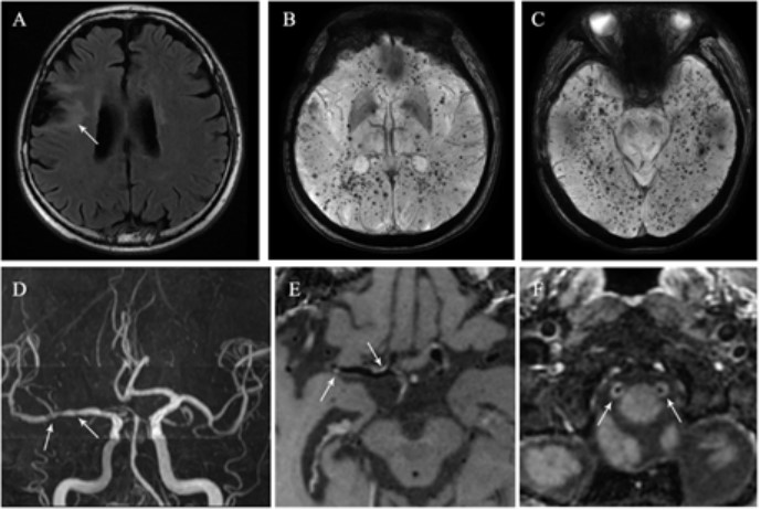
Figure 1. A
45-year-old man who had undergone radiation therapy after surgical resection of
frontal lobe glioma at 26 years old. (A) FLAIR imaging shows peri-lesion white
matter lesions. (B) and (C) SWI shows multiple cerebral microbleeds locating
widespread the brain. (D) MRA shows multiple intracranial arteries stenosis.
(E) Post-contrast 3D T1W HR-MRI shows eccentric plaques with moderate focal
enhancement (E, arrow) in right M1 segment of middle cerebral artery. (F)
Post-contrast 3D T1W HR-MRI shows prominent circle enhancement in bilateral V4
segment of vertebral arteries.
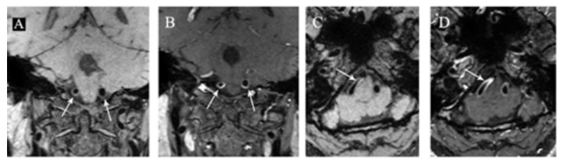
Figure 2. A
47-year-old woman who had undergone radiation therapy for pituitary adenoma at
36 years old. Pre-contrast (A) and post-contrast (B) 3D T1W HR-MRI show
concentric wall thickening (A, arrow) with prominent circle enhancement (B,
arrow) in bilateral V4 segment of vertebral arteries on coronal images.
Pre-contrast (C) and post-contrast (D) 3D T1W HR-MRI show track train
sign-concentric wall thickening (C, arrow) with prominent homogeneous
enhancement (D, arrow) in right V4 segment of vertebral artery on axial images.