Dongsuk Sung1, Peter A. Kottke2, Jason W. Allen1,3,4, Benjamin B. Risk5, Fadi Nahab4, Andrei G. Fedorov2,6, and Candace C. Fleischer1,3,6
1Department of Biomedical Engineering, Georgia Institute of Technology and Emory University, Atlanta, GA, United States, 2Woodruff School of Mechanical Engineering, Georgia Institute of Technology, Atlanta, GA, United States, 3Department of Radiology and Imaging Sciences, Emory University School of Medicine, Atlanta, GA, United States, 4Department of Neurology, Emory University School of Medicine, Atlanta, GA, United States, 5Department of Biostatistics and Bioinformatics, Emory University, Atlanta, GA, United States, 6Petit Institute for Bioengineering and Bioscience, Georgia Institute of Technology, Atlanta, GA, United States
1Department of Biomedical Engineering, Georgia Institute of Technology and Emory University, Atlanta, GA, United States, 2Woodruff School of Mechanical Engineering, Georgia Institute of Technology, Atlanta, GA, United States, 3Department of Radiology and Imaging Sciences, Emory University School of Medicine, Atlanta, GA, United States, 4Department of Neurology, Emory University School of Medicine, Atlanta, GA, United States, 5Department of Biostatistics and Bioinformatics, Emory University, Atlanta, GA, United States, 6Petit Institute for Bioengineering and Bioscience, Georgia Institute of Technology, Atlanta, GA, United States
A personalized MR-derived brain temperature model was developed using patient data. Simulated brain temperatures after right middle cerebral artery occlusion were highest in the ischemic penumbra followed by infarct core, contralateral healthy tissue, and ipsilateral healthy tissue.
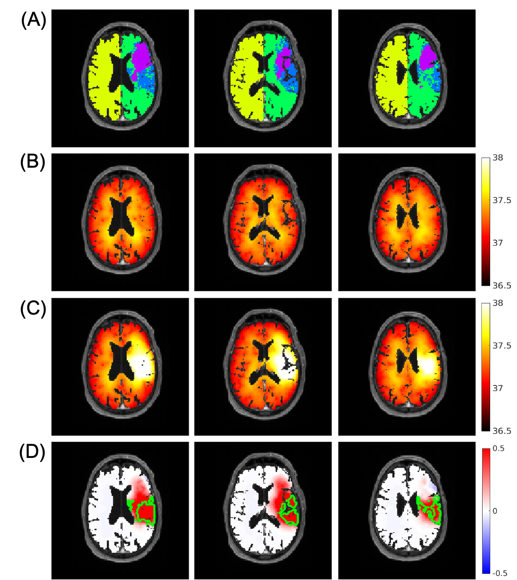
Figure 2. Representative images from a patient with ischemic stroke showing the ability of brain temperature to differentiate regions after ischemic stroke. (A) Tissue regions defined from MR images include infarct core (purple), ischemic penumbra (blue), ipsilateral (green), and contralateral (yellow) healthy tissue; (B) Simulated brain temperature map before occlusion; (C) Simulated brain temperature map after 100% occlusion; (D) Brain temperature difference map (post- minus pre-occlusion). The green boundaries are major continuous regions of ischemic penumbra.
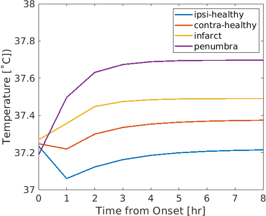
Figure 3. Predicted brain temperature evolution from onset to 8 hours after right middle cerebral artery occlusion in four regions: ipsilateral healthy tissue (ipsi-healthy), contralateral tissue (contra-healthy), infarct core (infarct), and ischemic penumbra (penumbra), where steady state brain temperatures were 37.21 ˚C, 37.37 ˚C, 37.49 ˚C, and 37.70 ˚C, respectively. These in silico results are consistent with previous in vivo studies and suggest that brain temperature may be used as a predictive biomarker to identify hemodynamically distinct regions.