Ryan Andrew Rava1,2, Kenneth V. Snyder2,3, Muhammad Waqas2,3, Elad I. Levy2,3, Jason M. Davies2,3, Adnan H. Siddiqui2,3, and Ciprian N. Ionita1,2,3
1Biomedical Engineering, University at Buffalo, Buffalo, NY, United States, 2Canon Stroke and Vascular Research Center, Buffalo, NY, United States, 3Neurosurgery, University at Buffalo, Buffalo, NY, United States
1Biomedical Engineering, University at Buffalo, Buffalo, NY, United States, 2Canon Stroke and Vascular Research Center, Buffalo, NY, United States, 3Neurosurgery, University at Buffalo, Buffalo, NY, United States
Two
neural networks were developed to segment penumbra using FLAIR and DWI. Metrics
comparing predictions with ground truth penumbra ((dual network, multi-input
network): Dice=(0.61, 0.61), PPV=(0.59, 0.64)) indicate a multi-input network
is more capable of segmenting penumbra.
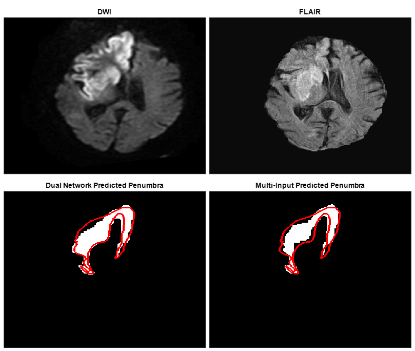
Figure
3: Top images indicate DWI and FLAIR slices fed into each network to segment
the penumbra seen in the bottom images. The dual network prediction indicates
the subtraction of FLAIR segmented infarct from DWI segmented ischemic tissue.
The multi-input prediction indicates predicted penumbra using both DWI and
FLAIR as network input and the red line is an outline of the ground truth
penumbra for this slice. Dual and multi-input Dice coefficients for this slice
are 0.83 and 0.78 respectively.
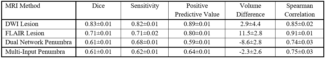
Table
1. Dice Coefficients, sensitivity, positive predictive values, infarct/ischemic
tissue/penumbra volume differences, and Spearman correlation coefficients, with
95% confidence intervals, for each network’s segmentation performance of either
ischemic tissue (DWI), infarct (FLAIR), or penumbra (Dual Network and
Multi-Input).