Shanmukha Srinivas1, Tara Retson1, Aaron Simon2, Marc Alley3, Shreyas Vasanawala3, Jona Hattangadi-Gluth2, Albert Hsiao1, and Nikdokht Farid1
1Department of Radiology, University of California San Diego, San Diego, CA, United States, 2Department of Radiation Medicine and Applied Sciences, University of California San Diego, San Diego, CA, United States, 3Department of Radiology, Stanford University, Stanford, CA, United States
1Department of Radiology, University of California San Diego, San Diego, CA, United States, 2Department of Radiation Medicine and Applied Sciences, University of California San Diego, San Diego, CA, United States, 3Department of Radiology, Stanford University, Stanford, CA, United States
Feeding arterial and draining venous blood flow, as measured on 4D flow MRI, decreased in cerebral AVMs after SRS. Pulsatility index increased within the feeding artery. Arterial circumference did not decrease significantly unlike AVM nidus volume and venous circumference.
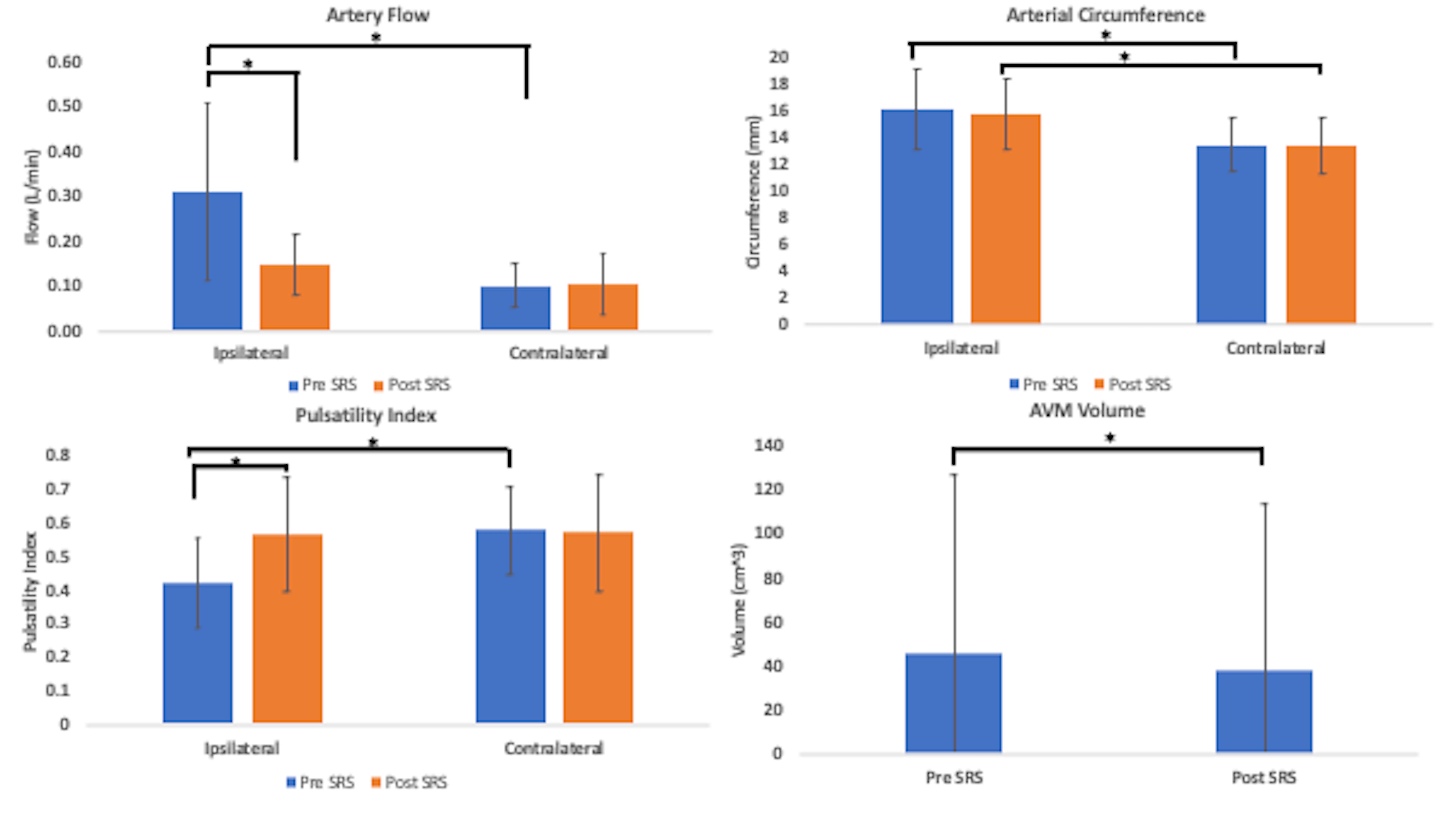
Bar graph of total changes in arterial flow, circumference, pulsatility index and AVM volume before and after SRS. Ipsilateral artery flow was significantly greater than contralateral artery flow pre-SRS and decreased significantly post-SRS. Ipsilateral artery circumference was greater than contralateral artery circumference before and after SRS. Ipsilateral artery pulsatility was lower than contralateral artery pulsatility prior to SRS and increased significantly after SRS. Volume significantly decreased after SRS.
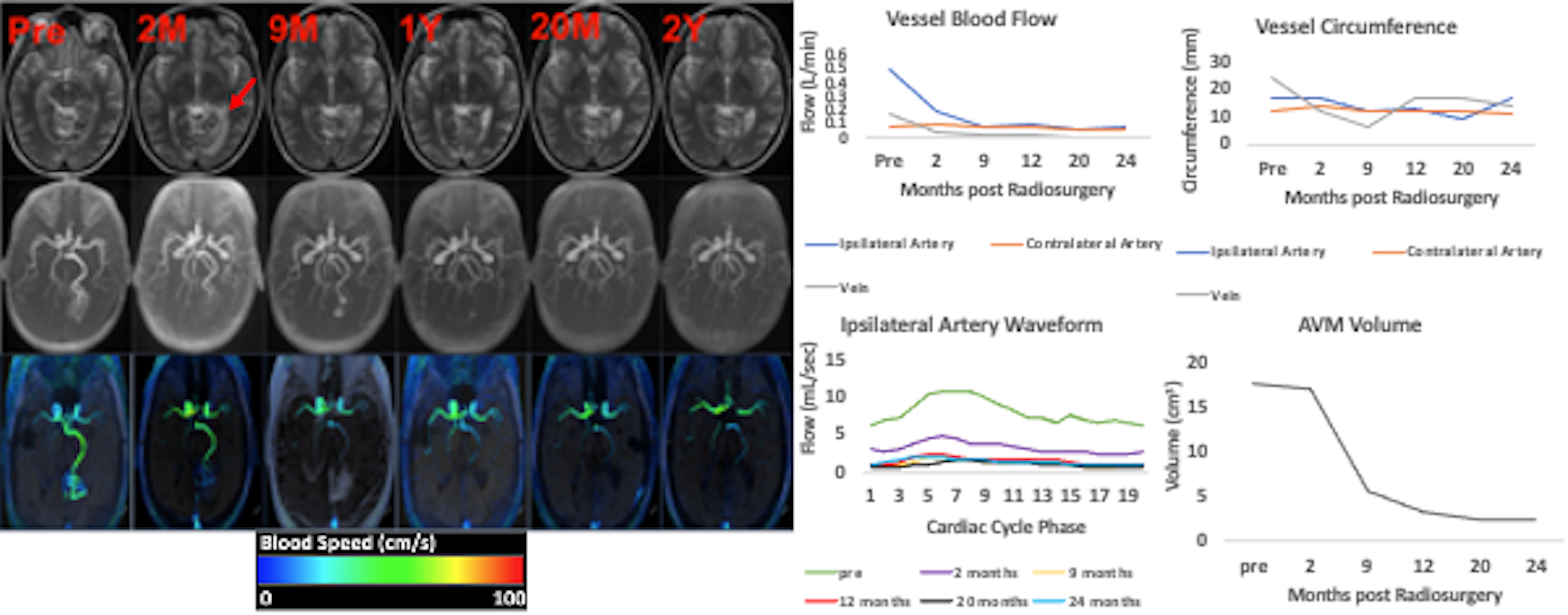
33 year old female with left posterior temporal-occipital AVM treated with a single-stage of SRS (1600 cGy). While T2W imaging (1A, G) and MRA (1B) demonstrate gradual decrease of the nidus from before SRS to 2 years after SRS, 4D Flow (1C, D, F) demonstrates a 61.2% reduction in feeding artery blood flow and 76.5% reduction in draining vein blood flow by 2 months, and normalization of feeding artery blood flow by 2 years. In comparison, there is only 2.3% reduction in feeding artery circumference with a more substantial reduction in draining vein circumference (50.6%) by 2 months (1E).