Cecilia Björnfot1, Anders Garpebring1, Sara Qvarlander1, Jan Malm2, Anders Eklund1, and Anders Wåhlin1,3
1Department of Radiation Sciences, Umeå University, Umeå, Sweden, 2Department of Clinical Sciences, Umeå University, Umeå, Sweden, 3Umeå Center for Functional Brain Imaging, Umeå University, Umeå, Sweden
1Department of Radiation Sciences, Umeå University, Umeå, Sweden, 2Department of Clinical Sciences, Umeå University, Umeå, Sweden, 3Umeå Center for Functional Brain Imaging, Umeå University, Umeå, Sweden
We present a 4D flow MRI method to estimate pulse wave velocity in the intracranial arterial tree. The method is shown to be stable in an internal consistency test, and of sufficient sensitivity to robustly detect age-related increases in intracranial pulse wave velocity
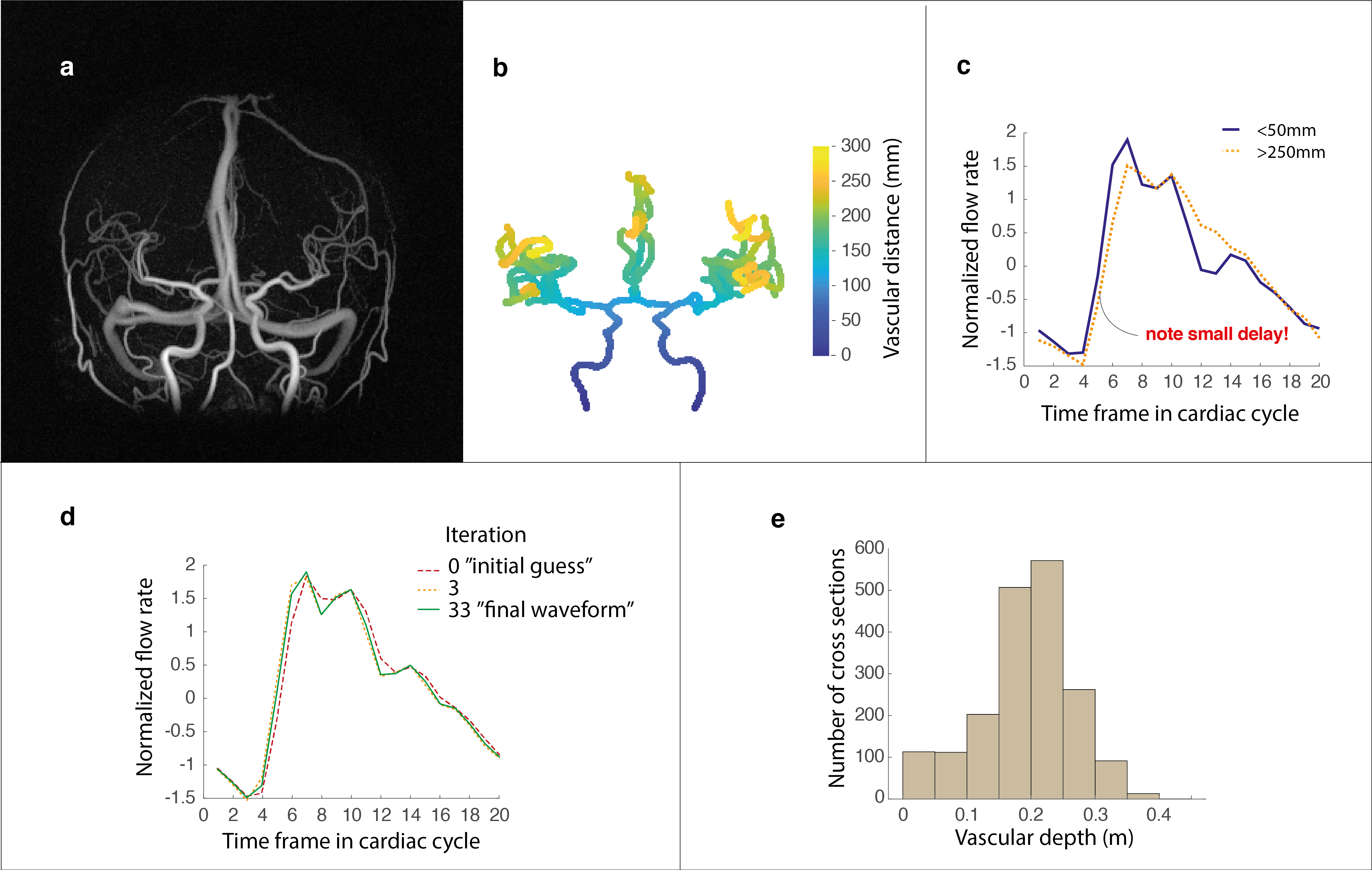
Figure 1. (a) Angiographic image and (b) a corresponding centerline representation with calculated vascular depth. (c) The average flow rate waveforms obtained in the most proximal (depth<50mm) as well as in the most distal aspects (depth>250mm) of the visible vasculature. (d) The initial guess, 3rd and 33rd iteration of the estimated arterial waveform obtained from the minimization process. (e) Histogram illustrating the average number of cross sections available as a function of depth in the vascular tree.