Piotr Radojewski1, Arun Joseph2,3,4, Gabriele Bonnano2,3,4, Tom Hilbert5,6,7, Tobias Kober5,7,8, Jan Gralla1, Roland Wiest1, and Pasquale Mordasini1
1Institute of Diagnostic and Interventional Neuroradiology, Bern University Hospital, Inselspital,, Bern, Switzerland, 22. Advanced Clinical Imaging Technology, Siemens Healthcare AG, Bern, Switzerland, 33. Translational Imaging Center, Sitem-Insel, Bern, Switzerland, 44. Departments of Radiology and Biomedical Research, University of Bern, Bern, Switzerland, 55. Advanced Clinical Imaging Technology, Siemens Healthcare AG, Lausanne, Switzerland, 66. Department of Radiology, , Lausanne University Hospital and University of Lausanne, Lausanne, Switzerland, 77. LTS5, École Polytechnique Fédérale de Lausanne (EPFL), Lausanne, Switzerland, 86. Department of Radiology, Lausanne University Hospital and University of Lausanne, Lausanne, Switzerland
1Institute of Diagnostic and Interventional Neuroradiology, Bern University Hospital, Inselspital,, Bern, Switzerland, 22. Advanced Clinical Imaging Technology, Siemens Healthcare AG, Bern, Switzerland, 33. Translational Imaging Center, Sitem-Insel, Bern, Switzerland, 44. Departments of Radiology and Biomedical Research, University of Bern, Bern, Switzerland, 55. Advanced Clinical Imaging Technology, Siemens Healthcare AG, Lausanne, Switzerland, 66. Department of Radiology, , Lausanne University Hospital and University of Lausanne, Lausanne, Switzerland, 77. LTS5, École Polytechnique Fédérale de Lausanne (EPFL), Lausanne, Switzerland, 86. Department of Radiology, Lausanne University Hospital and University of Lausanne, Lausanne, Switzerland
Integration of ultra-high-field MR neurovascular vessel
imaging at 7 Tesla into clinical routine is feasible providing higher spatial
resolution and contrast which may result in an increased diagnostic confidence.
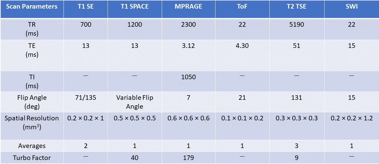
Scan parameters of T1 SE, T1 SPACE,
MPRAGE, ToF, T2 TSE, and SWI protocols used for the vessel wall imaging
measurements at 7T.
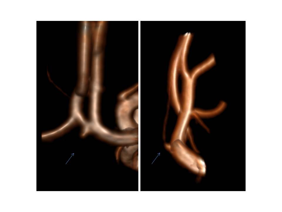
Anatomico-morphological characterization. 3D reconstruction of arterial ToF sequence at 3T
MRI (left panel) and 7T MRI (right panel) in a case of a suspected UIA. 7T revealed the
presence of an infundibulary branch mimicking UIA.