Niranjan Balu1, Wenjin Liu1, Zhensen Chen1, Anders Gould1, Dan S Hippe1, Li Chen1, Binbin Sui2, Mi Shen2, Peiyi Gao2, Thomas S Hatsukami1, and Chun Yuan1
1Radiology, University of Washington, Seattle, WA, United States, 2Beijing Tiantan Hospital, Beijing, China
1Radiology, University of Washington, Seattle, WA, United States, 2Beijing Tiantan Hospital, Beijing, China
Dependence of measurement reproducibility of intracranial
distal vessel length on TOF-MRA on protocol and scanner platform variation was
investigated. High reproducibility was
achieved suggesting feasibility of quantitative vessel length measurements for
multi-platform studies.
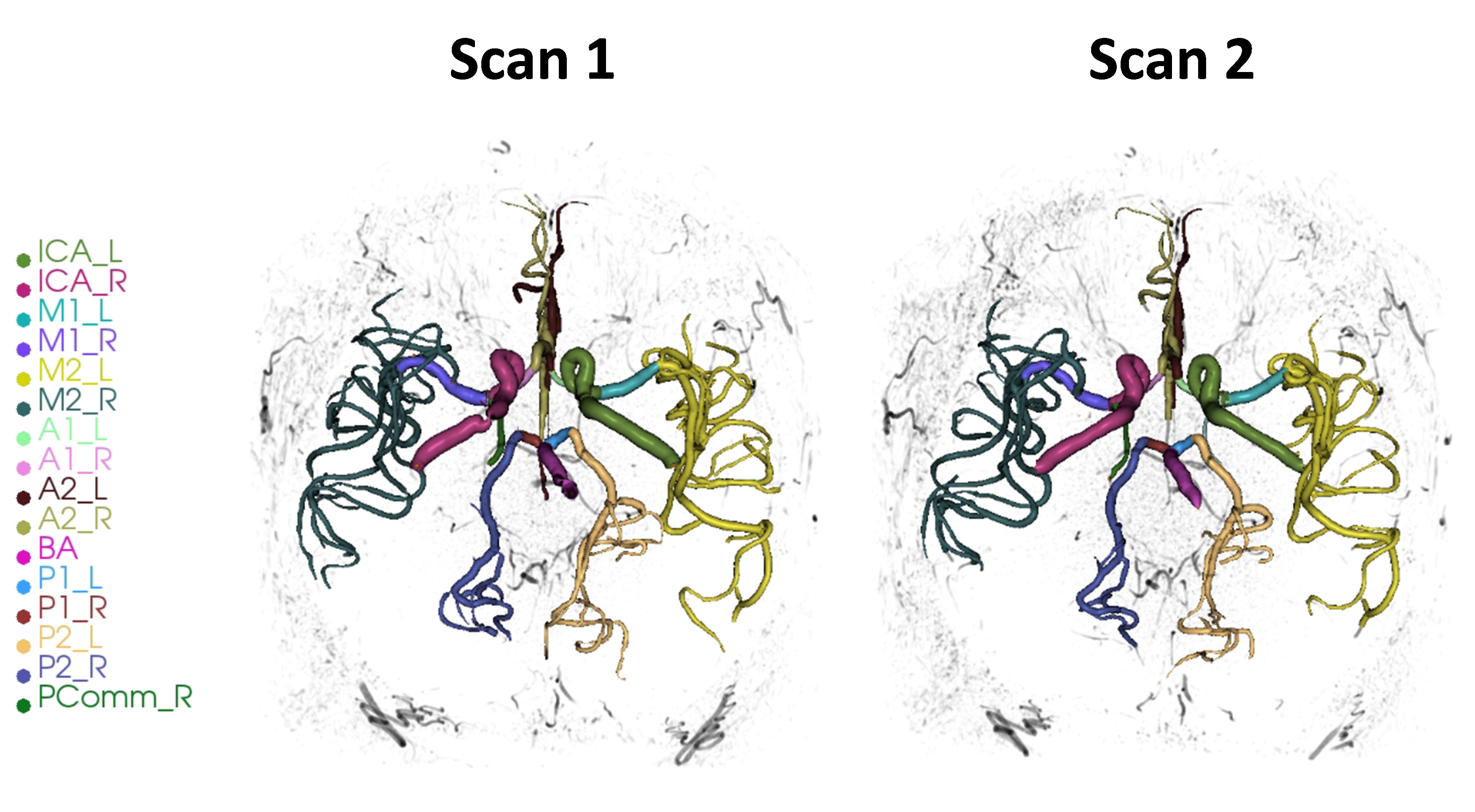
Figure 1: Segmented arteries by iCafe from two
Philips 3T TOF-MRA (S/I FOV 7.2cm) 2 weeks apart. The different colors indicate
different arteries as denoted by the labels (Left (denoted by suffix L) and
right (denoted by suffix R) sided ICA- internal carotid artery, M1/A2 – Middle
cerebral artery M1, M2 branches, A1/A2 - Anterior cerebral artery A1, A2
branches), BA – Basilar artery, P1/P2 – posterior cerebral artery P1, P2
branches, PComm – posterior communicating artery). Quantitative artery length
measurements can be derived based on the centerlines from these iCafe
segmentations.
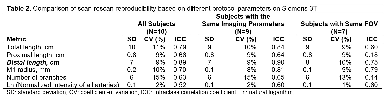
Table 2. Comparison of
scan-rescan reproducibility based on different protocol parameters on Siemens
3T