Adam E. Goldman-Yassen1, Eytan Raz2, Anna Derman2, Ahrya Derakhshani3, and Seena Dehkharghani2,4
1Department of Radiology, Children's Healthcare of Atlanta, Atlanta, GA, United States, 2Department of Radiology, New York University Langone Medical Center, New York, NY, United States, 3Department of Radiology, UCLA Health, Los Angeles, CA, United States, 4Department of Neurology, New York University Langone Medical Center, New York, NY, United States
1Department of Radiology, Children's Healthcare of Atlanta, Atlanta, GA, United States, 2Department of Radiology, New York University Langone Medical Center, New York, NY, United States, 3Department of Radiology, UCLA Health, Los Angeles, CA, United States, 4Department of Neurology, New York University Langone Medical Center, New York, NY, United States
High-resolution black-blood vessel wall imaging optimized with robust flow suppression offers reproducible, reliable, and non-invasive evaluation of flow diversion treated aneurysms, with superior overall classification accuracy relative to conventional TOF or dynamic MRA.
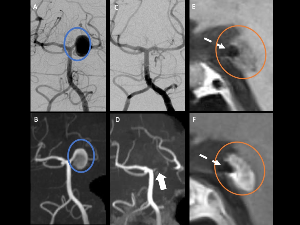
DSA (A) and TOF MRA (B) show an aneurysm at the left SCA and PCA junction (blue circle). Followup DSA (C) after basilar/left PCA flow-diverting stent placement demonstrates complete occlusion. MRA demonstrates questionable flow-related enhancement not easily discriminated from T1 shine through from organizing clot (D). Pre (E) and postcontrast (F) VWI demonstrate preserved high-resolution black blood angiography across the stented PCA segment (dashed arrow). Organizing clot within the aneurysm sac (orange circle) is noted, likely fed through a vasa vasorum, without flow voids.
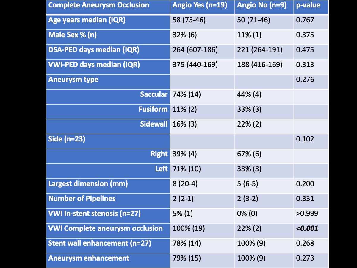
Comparison of patient characteristics and vessel wall imaging findings between subjects with and without complete aneurysm occlusion of followup angiogram after flow diverter placement.