1The First Hospital of JiLin University, Changchun City,Jilin Province, China, 2Philips healthcare, Guangzhou City,Guangdong Province, China
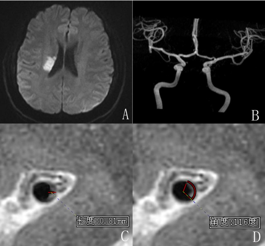
Figure 2:Hyper signal could be seen in the right corona radiata,so the MCA plaque is a symptomatic plaque(A).
MRA showed the mild stenosis of the right middle cerebral artery.(B)
The maximum wall thickness as measured using the MRI-PlaqueView software at the leision slice of the right middle artery was 0.81 mm.(C)
The plaque distribution range measured using RadiAnt DICOM Viewer software at the leison slice of the right middle artery was 116°.(D)

Figure 3:Hyper signal could be seen in the left cerebral peduncle, so the plaque was a symptomatic plaque(A).
The plaque distribution range at the leision slice of BA was 136°.(B)
The maximum wall thickness and the minimum wall thickness for the lesion slice were 2.07 mm and 0.41 mmrespectively (C and D)