So Yeon Won1, Jihoon Cha1, Hyun Seok Choi1, Young Dae Kim2, Hyo Suk Nam2, Ji Hoe Heo2, and Seung-Koo Lee1
1Radiology, Severance Hospital, Seoul, Korea, Republic of, 2Neurology, Severance Hospital, Seoul, Korea, Republic of
1Radiology, Severance Hospital, Seoul, Korea, Republic of, 2Neurology, Severance Hospital, Seoul, Korea, Republic of
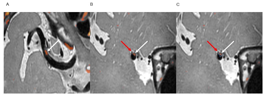
In patients with BOD, the plaque margin was closer to perforator
orifice with less stenosis and enhancement than patients with artery to artery
embolism.
Figure
2. Representative case with BOD. A. Longitudinal plane of MCA with fusion
image (PD+T1CE). B. Cross-sectional image where perforator arouses.
Perforator (red arrow), plaque (white arrow) C. Plaque distribution was
expressed as an angle. Perforator was located at margin of the plaque.
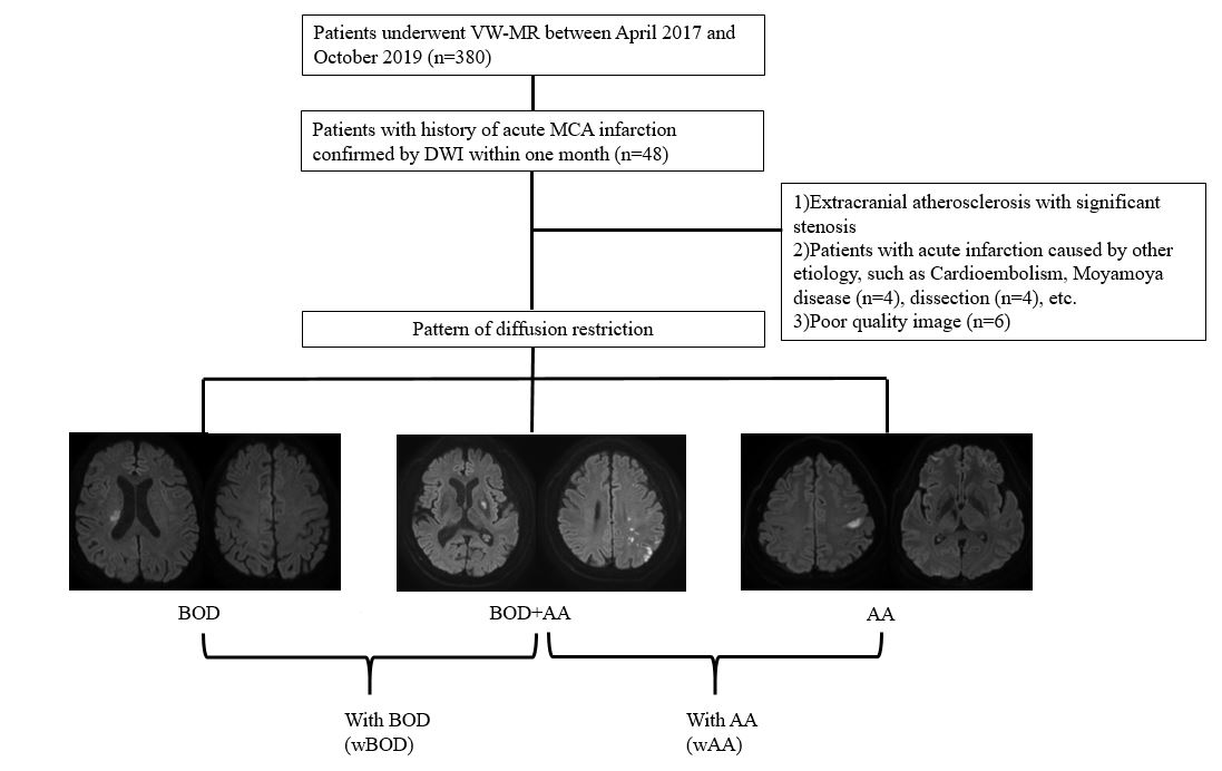
Figure 1.Diagram of patient selection.