Hongwei Zhou1, Derui Kong1, and Tianjing Zhang2
1The First Hospital of JiLin University, Changchun City,Jilin Province, China, 2Philips healthcare, Guangzhou City,Guangdong Province, China
1The First Hospital of JiLin University, Changchun City,Jilin Province, China, 2Philips healthcare, Guangzhou City,Guangdong Province, China
The purpose of our study was to summarize the typical imaging performance of PACNS and evaluate the value of 3D- VW-MRI sequence in demonstrating the detailed information in detection, diagnosis, evaluation, and follow-up for PACNS.
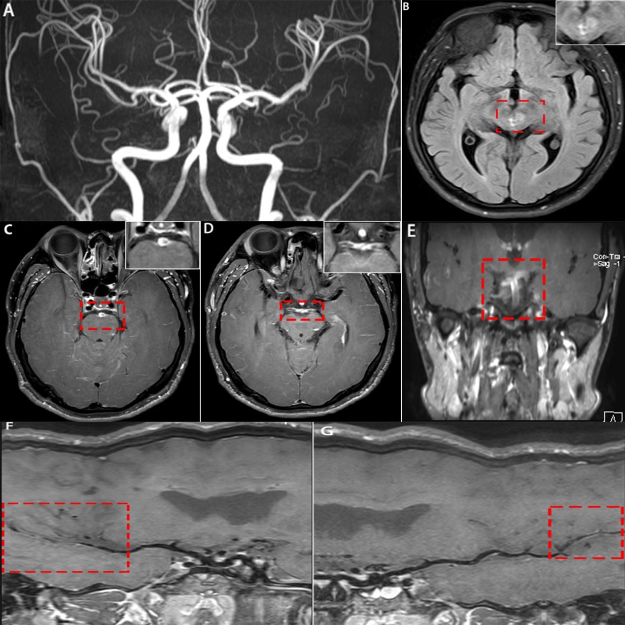
Figure 1:(A): MRA demonstrated smooth wall and no obvious stenosis. (B): Head MRI showed abnormal signals in the left oculomotor nucleus. (C-E): 3D VW-MRI showed unevenly thickening and abnormal enhancement of basilar artery,bilateral posterior cerebral artery and middle cerebral artery. (F-G):Curved plannar reconstruction by 3D-VW-MRI showed the left and right middle cerebral artery had no apparent enhancement after 4 months therapy, while the vessel wall was getting thickness.
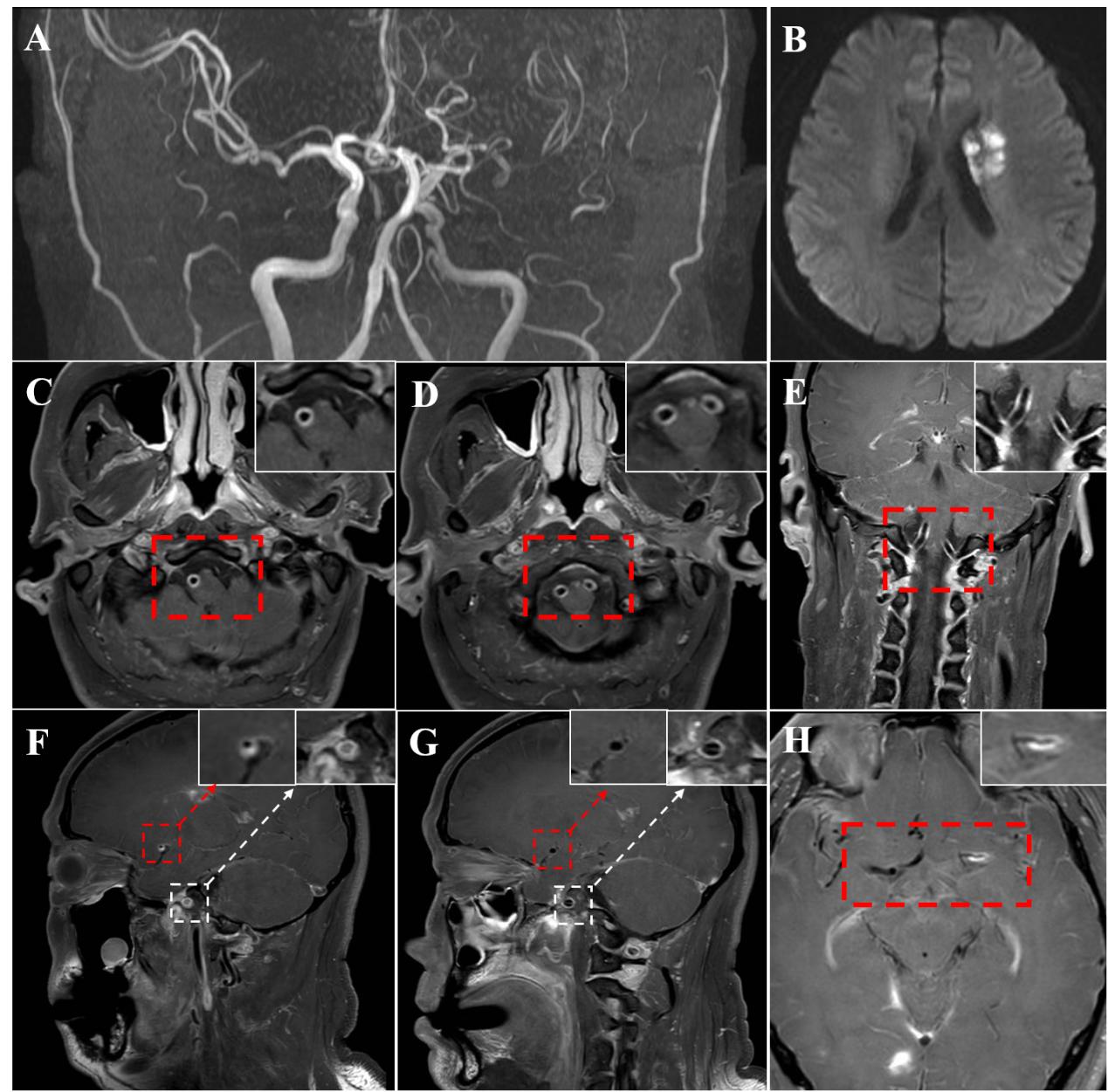
Figure 2:(A):MRA revealed multiple stenosis of both the anterior circulation and the posterior circulation. (B~H)3D VW-MRI showed smooth, concentric arterial wall thickening and enhancement of bilateral vertebral artery, the left internal carotid artery and middle cerebral artery.