Mengjiao Wei1, Yang Gao1, Qiong Wu1, Shaoyu Wang2, and Huapeng Zhang2
1Department of Radiology, Affiliated Hospital of Inner Mongolia Medical University, Hohhot, China, 2MR Scientific Marketing, Siemens Healthineers, Shanghai, China
1Department of Radiology, Affiliated Hospital of Inner Mongolia Medical University, Hohhot, China, 2MR Scientific Marketing, Siemens Healthineers, Shanghai, China
The maximum wall thickness, the maximum lipid-rich necrotic core percentage and the CAS score can accurately predict the risk of stroke and provide evidence for diagnosis and treatment.
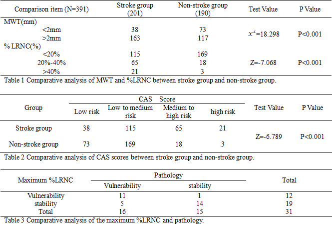
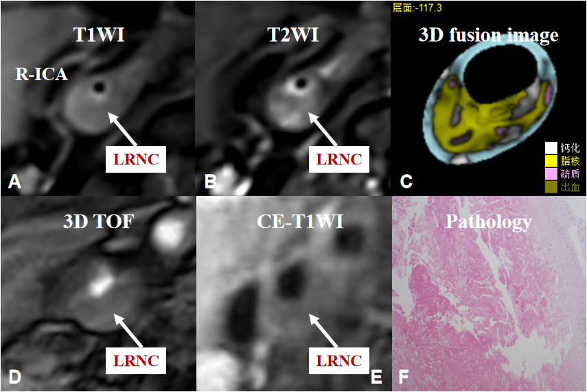
Figure 1. Typical images of the right internal carotid artery(R-ICA). (A)(B)(D)HR-VWI shows an isointensity plaque on T1WI, T2WI and TOF sequences on the right internal carotid artery wall (arrow) without postgadolinium enhancement(E). (C)3D fusion image shows that maximum lipid-rich necrotic core percentage > 40%. (F)Pathological result shows that a large amount of lipid components in the plaque.