Haonan Zhang1, Qingwei Song1, Jiazheng Wang2, Peng Sun2, Renwang Wang1, Nan Zhang1, and Ailian Liu1
1Department of Radiology, the First Affiliated Hospital of Dalian Medical University, Dalian, China, 2PHILIPS——Philips Healthcare, beijing, China
1Department of Radiology, the First Affiliated Hospital of Dalian Medical University, Dalian, China, 2PHILIPS——Philips Healthcare, beijing, China
In
this study, we demonstrated that a large CS acceleration factor will degrade the
image quality of 3D-TOF-MRA for cervical vessels significantly. CS factor of 6 is
recommended for the compromise between imaging time and image quality of clinical
3D-TOF carotid MRA.
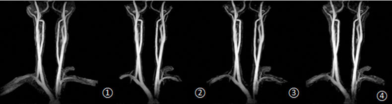
Figure
2. Reconstruction of cervical vessels, from
left to right: default, CS4-CS8.
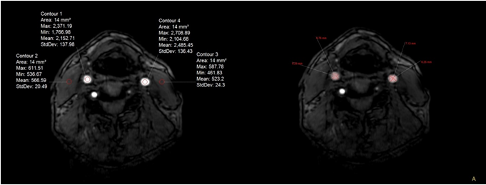
Figure
1. Male, 68 years old. Use ROI to measure SI and SD on both sides. The measured
left blood vessel SI value was 2485.45, muscle SI value was 523.2, and muscle
SD value was 24.3. The measured right blood vessel SI value was 2152.71, muscle
SI value 566.59, and muscle SD value 20.49. The largest and smallest diameters
of internal carotid arteries on both sides were measured. The largest diameter
measured on the right side is 7.13mm, and the smallest diameter is 6.25mm. The
maximum diameter on the left is 6.76mm and the minimum diameter is 6.25mm.