Peter J Lally1, Matthew Grech-Sollars2,3, Joely Smith3,4, Ben Statton5, Paul M Matthews1,6, Karla L Miller7, and Neal K Bangerter4
1Department of Brain Sciences, Imperial College London, London, United Kingdom, 2Department of Surgery and Cancer, Imperial College London, London, United Kingdom, 3Department of Imaging, Imperial College Healthcare NHS Trust, London, United Kingdom, 4Department of Bioengineering, Imperial College London, London, United Kingdom, 5MRC London Institute of Medical Sciences, London, United Kingdom, 6UK Dementia Research Institute Centre at Imperial College, London, United Kingdom, 7Wellcome Centre for Integrative Neuroimaging, Nuffield Department of Clinical Neurosciences, University of Oxford, Oxford, United Kingdom
1Department of Brain Sciences, Imperial College London, London, United Kingdom, 2Department of Surgery and Cancer, Imperial College London, London, United Kingdom, 3Department of Imaging, Imperial College Healthcare NHS Trust, London, United Kingdom, 4Department of Bioengineering, Imperial College London, London, United Kingdom, 5MRC London Institute of Medical Sciences, London, United Kingdom, 6UK Dementia Research Institute Centre at Imperial College, London, United Kingdom, 7Wellcome Centre for Integrative Neuroimaging, Nuffield Department of Clinical Neurosciences, University of Oxford, Oxford, United Kingdom
Here we describe a super-resolution 3D T2
relaxometry approach using an unbalanced SSFP acquisition with very low flip
angle RF pulses (α≤1°), and apply this in a phantom. The proposed approach provides new options for high-resolution,
low-SAR T2 relaxometry experiments in a range of tissues.
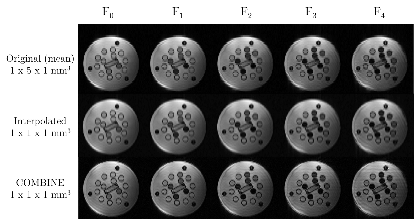
Figure 4: Top) Mean
low-resolution images (1x5x1mm3) for each of the different F-states
in the T2-COMBINE acquisition (TR=8ms, TE=4ms, α=1°).
Centre) Bicubic interpolation of the mean
low-resolution images (top) to a nominal voxel size of 1mm isotropic.
Bottom) Corresponding COMBINE reconstruction to a nominal voxel size of
1mm isotropic.
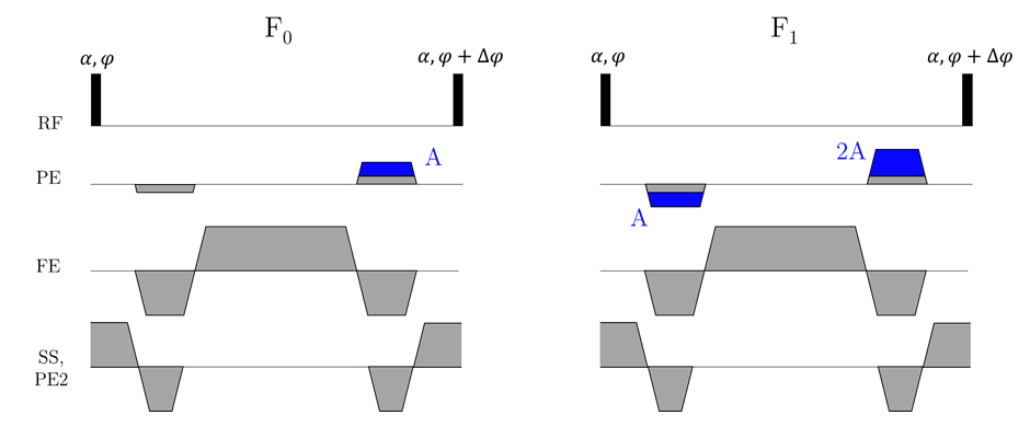
Figure 2: Schematic of the
pulse sequence for the T2-COMBINE experiment. Higher order F-states
are reached by adding integer increments of the unbalanced gradient area
(denoted A) either side of the readout.