Zoe Doyle1, Daehyun Yoon 1, Philip Lee1, Brian Hargreaves1, and Kathryn Stevens1
1Stanford University, Stanford, CA, United States
1Stanford University, Stanford, CA, United States
Our fast isotropic 3D MSI technique can facilitate the improved visualization of post-operative complications at 3T MRI by enabling the generation of clinically useful reformats in arbitrary imaging planes.
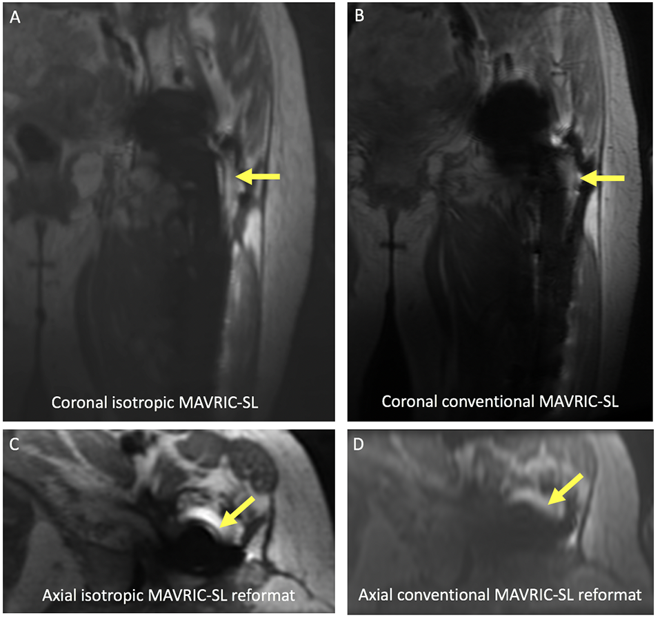
Fig 2. (A,B) Coronal and (C,D) axial reformatted images of a painful left revision total hip replacement in a 61-year-old female demonstrating abnormal signal adjacent to the lateral aspect of the femoral component on isotropic images (arrow). However, this abnormality cannot be seen on conventional MAVRIC SL images, despite the overall image quality being graded higher. Signal abnormality corresponded to fluid on inversion recovery sequences, confirming loosening (not shown).
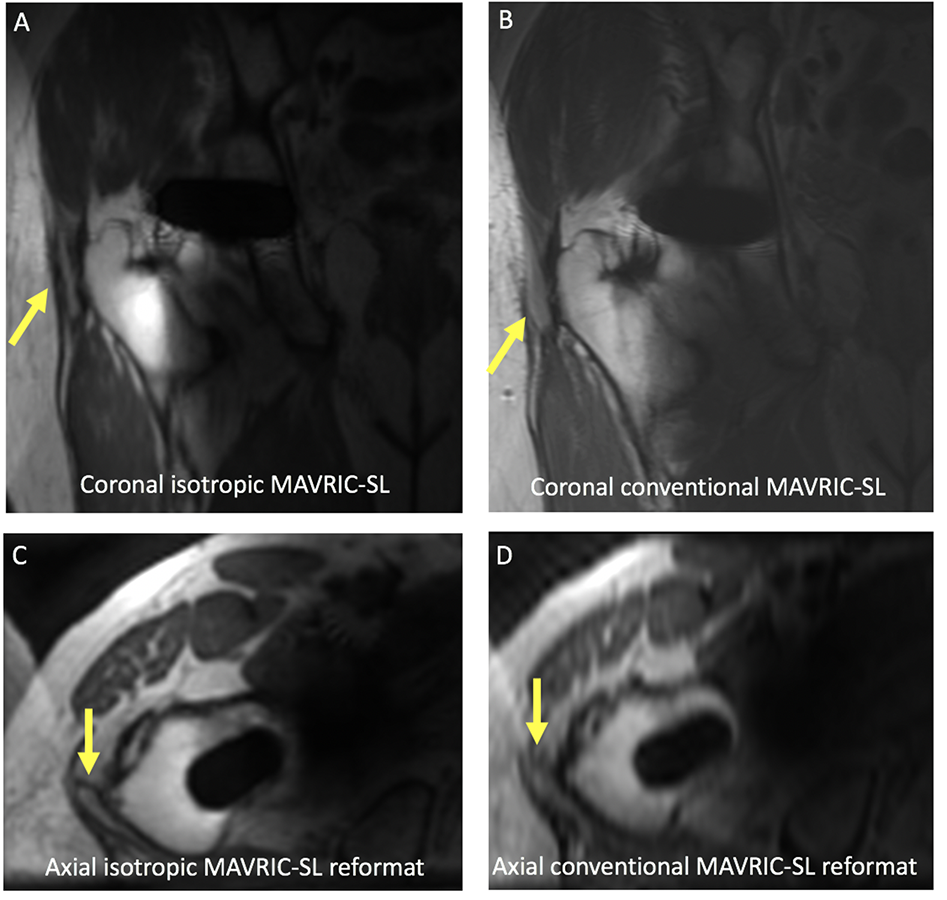
Fig 3. (A,B) Coronal and (C,D) axial reformatted images of a painful right total hip replacement in a 73-year-old male. A discrete trochanteric fluid collection is present (arrow), but is less well seen on conventional MAVRIC SL, particularly along the superior margin. Axial reformatted images confirm the presence of a trochanteric fluid collection, but the collection is less well seen on an axial reformatted conventional MAVRIC SL image (arrow).