Milena Capiglioni1, Claus Kiefer2, and Roland Wiest1
1Institute for Diagnostic and Interventional Neuroradiology, Support Center for Advanced Neuroimaging (SCAN), University of Bern, Bern, Switzerland, 2Institute for Diagnostic and Interventional Neuroradiology, Support Center for Advanced Neuroimaging (SCAN), Inselspital, Bern, Bern, Switzerland
1Institute for Diagnostic and Interventional Neuroradiology, Support Center for Advanced Neuroimaging (SCAN), University of Bern, Bern, Switzerland, 2Institute for Diagnostic and Interventional Neuroradiology, Support Center for Advanced Neuroimaging (SCAN), Inselspital, Bern, Bern, Switzerland
We detected oscillating fields in the nT range using a new Spin lock based contrast. This study takes a further step towards the use of neuronal current imaging in clinical applications.

Figure 5: Double resonance effect at two different frequencies of the Spin locking (a) 120 Hz and (b) 240 Hz. For both images, the frequency of the applied current was set at 120 Hz and the amplitude was calculated by the Biot-Savart law to be approximately 10 nT. To facilitate the visualization of the results, the rest of the deconvolution between SL on and SL off is displayed.
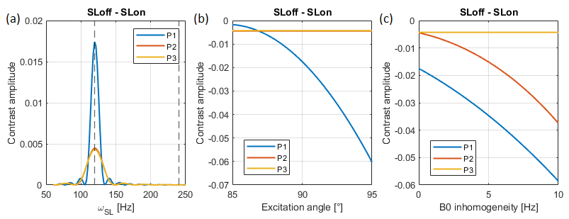
Figure 3: Bloch simulation of the behaviour of the three SL approaches showed in Figure 1. a) Contrast as a function of the SL frequency for homogeneous fields. b) Contrast in resonance (120 Hz) for the three approaches as a function of the excitation angle, where the variation is induced by B1 inhomogeneity. c) Contrast in resonance (120 Hz) for the three approaches as a function of the off resonant frequency induced by B0 inhomogeneity.