Sydney Nicole Williams1, Iulius Dragonu2, Patrick Liebig3, and David A. Porter1
1Imaging Centre of Excellence, University of Glasgow, Glasgow, United Kingdom, 2Siemens Healthcare Ltd., Frimley, United Kingdom, 3Siemens Healthineers, Erlangen, Germany
1Imaging Centre of Excellence, University of Glasgow, Glasgow, United Kingdom, 2Siemens Healthcare Ltd., Frimley, United Kingdom, 3Siemens Healthineers, Erlangen, Germany
We demonstrate slice-by-slice pTx shimming in a multi-slice readout-segmented diffusion-weighted sequence at 7 tesla. Slice-by-slice shimming is simple and efficient while greatly improving flip-angle homogeneity compared to volumetric-shimming and single-transmit.
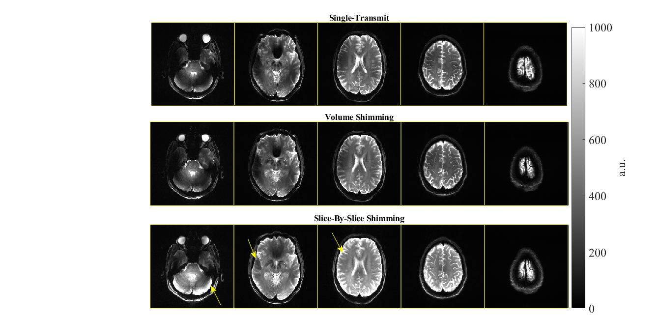
Figure 4. Experimental readout-segmented 2D images in a healthy subject: Top row) single-transmit, Middle row) volumetric-B1+ shimming, and Bottom row) slice-by-slice B1+ shimming. The arrows highlight a few areas where slice-by-slice shimming mitigates the B1+ inhomogeneity particularly well compared to single-transmit and volumetric-shimming. Meanwhile, there is a global improvement in the column 4 slice. Each column of slice images are windowed the same.
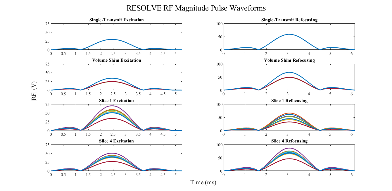
Figure 2. Magnitude RF waveforms in volts for 8 transmit channels, excitation pulse on the left and refocusing pulse on the right. Top row) single-transmit waveforms (all channels same magnitude with incremental 45° phase offset); Second row) volumetric-shim waveforms (in this case, the shimmed solution alternated between two magnitude values on all channels); Third row) slice 1 shim waveforms; Fourth row) slice 4 shim waveforms.