Kyle Jeong1, Noel Carlson2, John Rose3, and Eun-Kee Jeong4
1Utah Center for Advanced Imaging Research, University of Utah, Salt Lake City, UT, United States, 2NeuroImunology and NeuroVirology, University of Utah, Salt Lake City, UT, United States, 3Neurology, University of Utah, Salt Lake City, UT, United States, 4Radiology and Imaging Sciences, University of Utah, Salt Lake City, UT, United States
1Utah Center for Advanced Imaging Research, University of Utah, Salt Lake City, UT, United States, 2NeuroImunology and NeuroVirology, University of Utah, Salt Lake City, UT, United States, 3Neurology, University of Utah, Salt Lake City, UT, United States, 4Radiology and Imaging Sciences, University of Utah, Salt Lake City, UT, United States
DWSE and DWSTE were successfully combined for ultrahigh-b DWI of CSC imaging. The combined data set provides reliable signal-b curves for further quantitative analysis. The imaging time was only 6 min for 21 slices, bmax of 9781 and 3240 s/mm2 with 13 and 7 b-values for rDWI and aDWI, respectively.
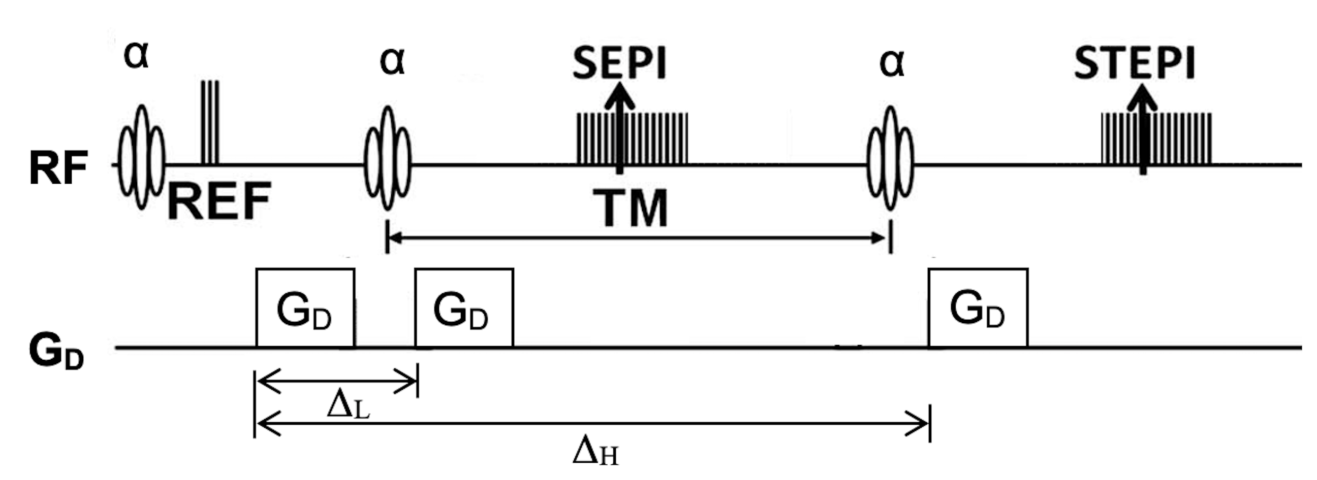
Fig. 1. Pulse sequence diagram of DW-SESTE with the flipangle α and amplitude GD and duration δ of the diffusion gradients.
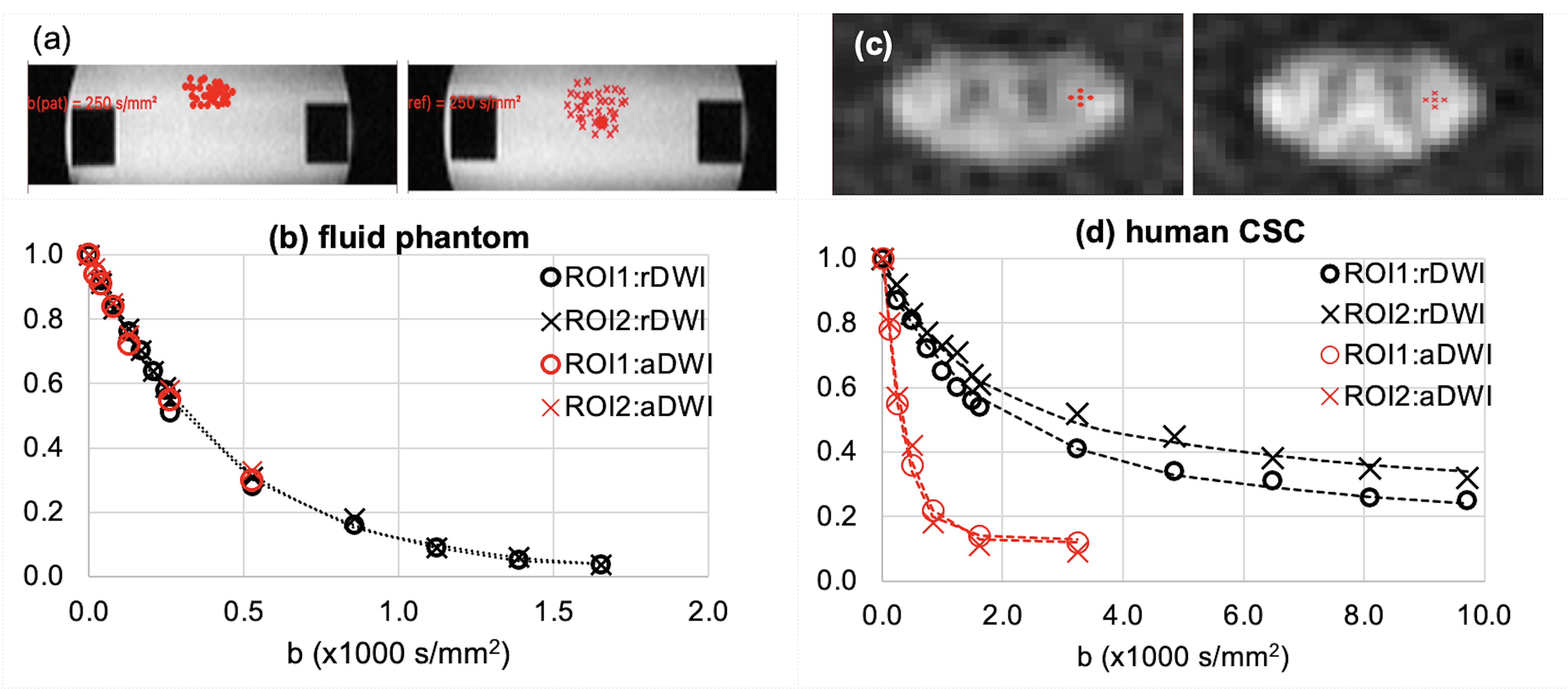
Fig. 2. (a, c) ROI (red dots and crosses) and (b, d) signal-b curves of UHb-DWI. Signal-b curves were fit to a single-exponential function for the phantom data in (b) and human aDWI data (red O, X)) in (d), and a double-exponential function for human rDWI data (black O, X) in (d).