Yajie Wang1, Yishi Wang2, Haikun Qi3, Rui Guo4, Huiyu Qiao1, Dongyue Si1, and Huijun Chen1
1Center for Biomedical Imaging Research, Department of Biomedical Engineering, School of Medicine, Tsinghua University, Beijing, China, 2Philips Healthcare, Beijing, China, 3School of Biomedical Engineering and Imaging Sciences, King's College London, London, United Kingdom, 4Department of Medicine, Beth Israel Deaconess Medical Center and Harvard Medical School, Boston, MA, United States
1Center for Biomedical Imaging Research, Department of Biomedical Engineering, School of Medicine, Tsinghua University, Beijing, China, 2Philips Healthcare, Beijing, China, 3School of Biomedical Engineering and Imaging Sciences, King's College London, London, United Kingdom, 4Department of Medicine, Beth Israel Deaconess Medical Center and Harvard Medical School, Boston, MA, United States
A
new quantitative technique using combined single- and multi-echo 3D golden angle
radial acquisition for simultaneous T1, T2 and T2* mapping of the carotid
plaque has been demonstrated to have good quantitative accuracy and in-vivo
feasibility.
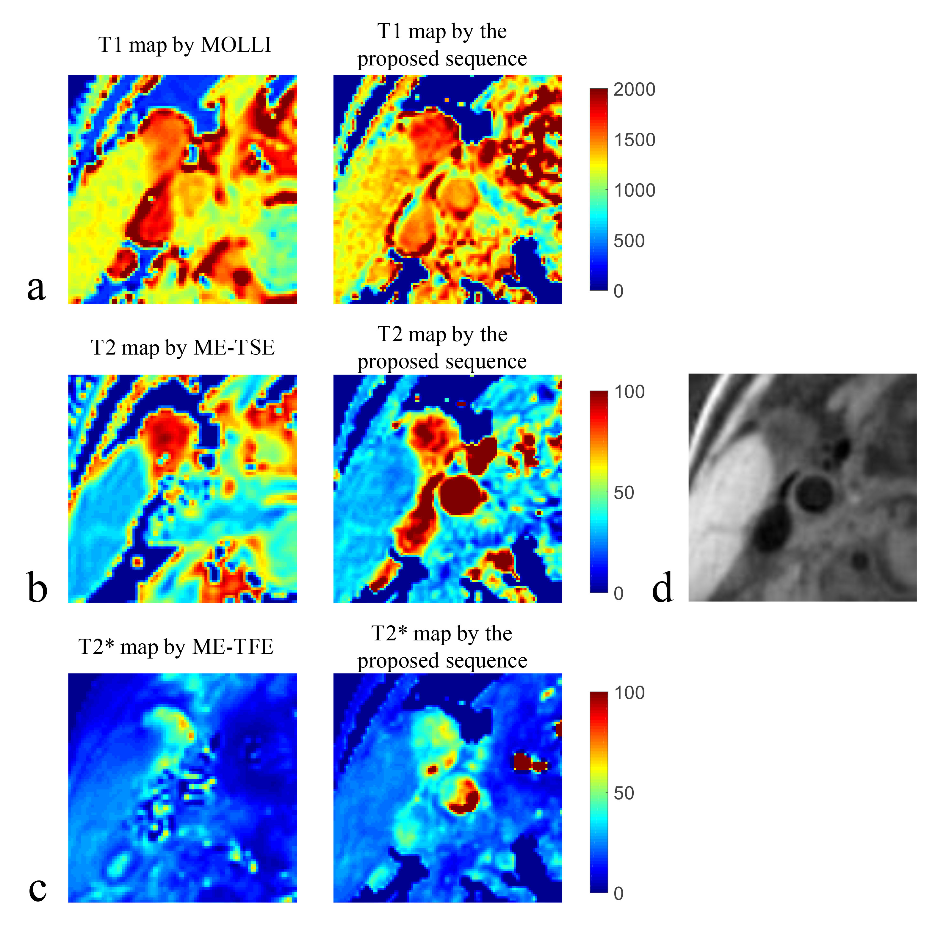
Figure
3. Quantitative mapping results of one healthy volunteer (male, 30 years). (a)
T1 maps estimated by modified Look-Locker inversion recovery
(MOLLI) and
the proposed sequence; (b) T2
maps
estimated by multi-echo
tubor spin
echo (ME-TSE)
and the proposed sequence;
(c) T2* maps
estimated by multi-echo
turbo
field
echo (ME-TFE)
and the proposed sequence;
(d) vessel wall image reconstructed from the proposed sequence.
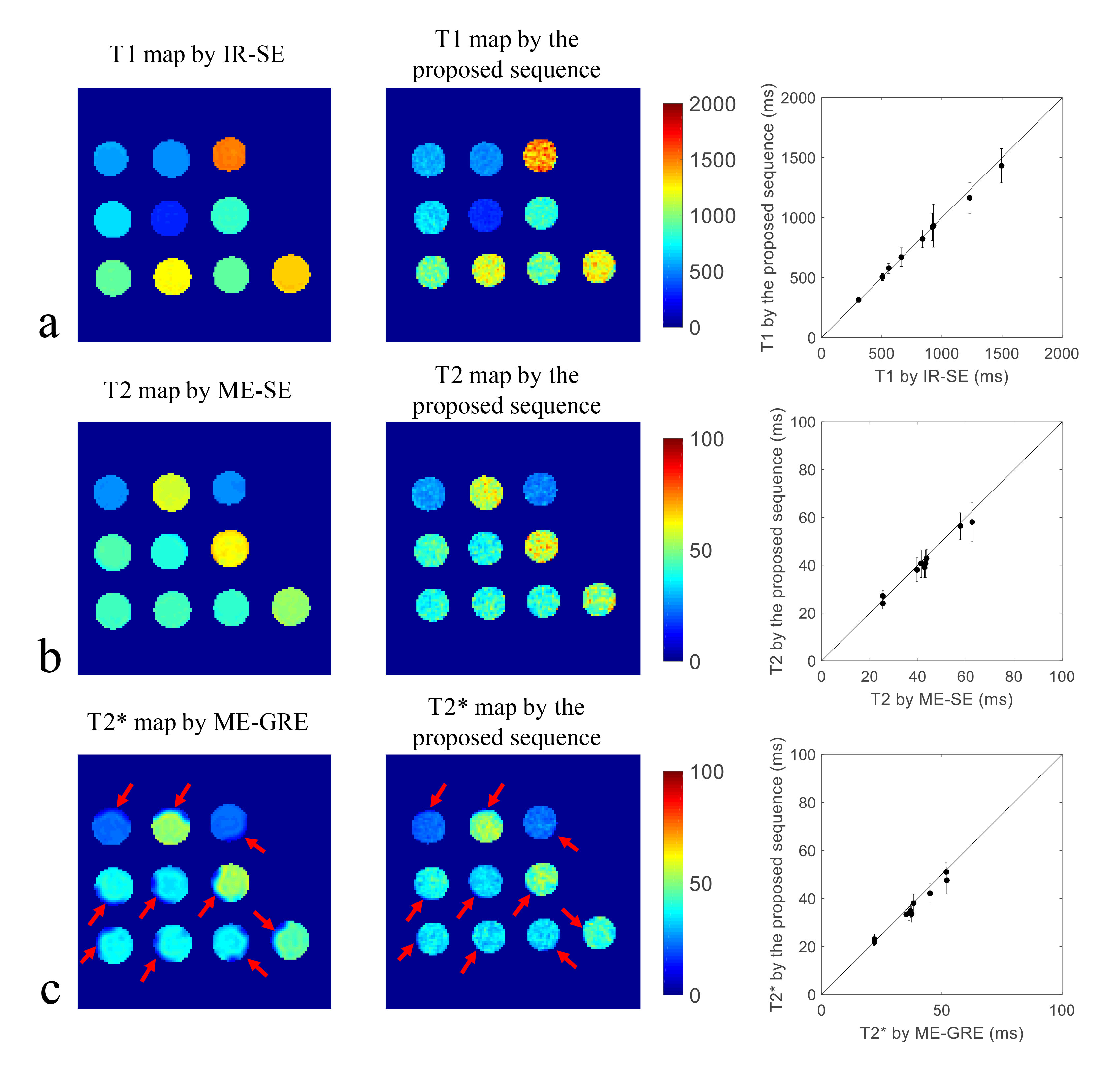
Figure
2. Quantitative mapping results of the
phantom. (a) T1 maps estimated
by
inversion
recovery spin echo (IR-SE)
and the proposed sequence with quantitative comparison (mean and SD); (b)
T2
maps
estimated
by
multi-echo
spin echo (ME-SE) and the proposed sequence with quantitative comparison (mean
and SD); (c)
T2*
maps
estimated
by
multi-echo
gradient echo (ME-GRE) and the proposed sequence with quantitative comparison
(mean and SD).