Serdest Demir1, Bryan Clifford2, Thorsten Feiweier3, Tom Hilbert4, Zahra Hosseini5, Augusto Lio Goncalves Filho1, Azadeh Tabari1, Wei-Ching Lo2, Maria Gabriela Figueiro Longo1, Michael Lev1, Pamela Schaefer1, Otto Rapalino1, Kawin Setsompop6, Berkin Bilgic6, Stephen Cauley6, Susie Huang1, and John Conklin1
1Radiology, Massachusetts General Hospital, Boston, MA, United States, 2Siemens Medical Solutions, Boston, MA, United States, 3Siemens Healthcare GmbH, Erlangen, Germany, 4Siemens Healthcare AG, Lausanne, Switzerland, 5Siemens Medical Solutions, Atlanta, GA, United States, 6Radiology, A. A. Martinos Center for Biomedical Imaging, Massachusetts General Hospital, Boston, MA, United States
1Radiology, Massachusetts General Hospital, Boston, MA, United States, 2Siemens Medical Solutions, Boston, MA, United States, 3Siemens Healthcare GmbH, Erlangen, Germany, 4Siemens Healthcare AG, Lausanne, Switzerland, 5Siemens Medical Solutions, Atlanta, GA, United States, 6Radiology, A. A. Martinos Center for Biomedical Imaging, Massachusetts General Hospital, Boston, MA, United States
EPI FLAIR images have lower
tissue contrast than TSE FLAIR images due to the absence of significant MT
effects. We demonstrate that an optimized MT-prepared EPI FLAIR acquisition can
achieve equivalent tissue contrast to conventional TSE FLAIR images.
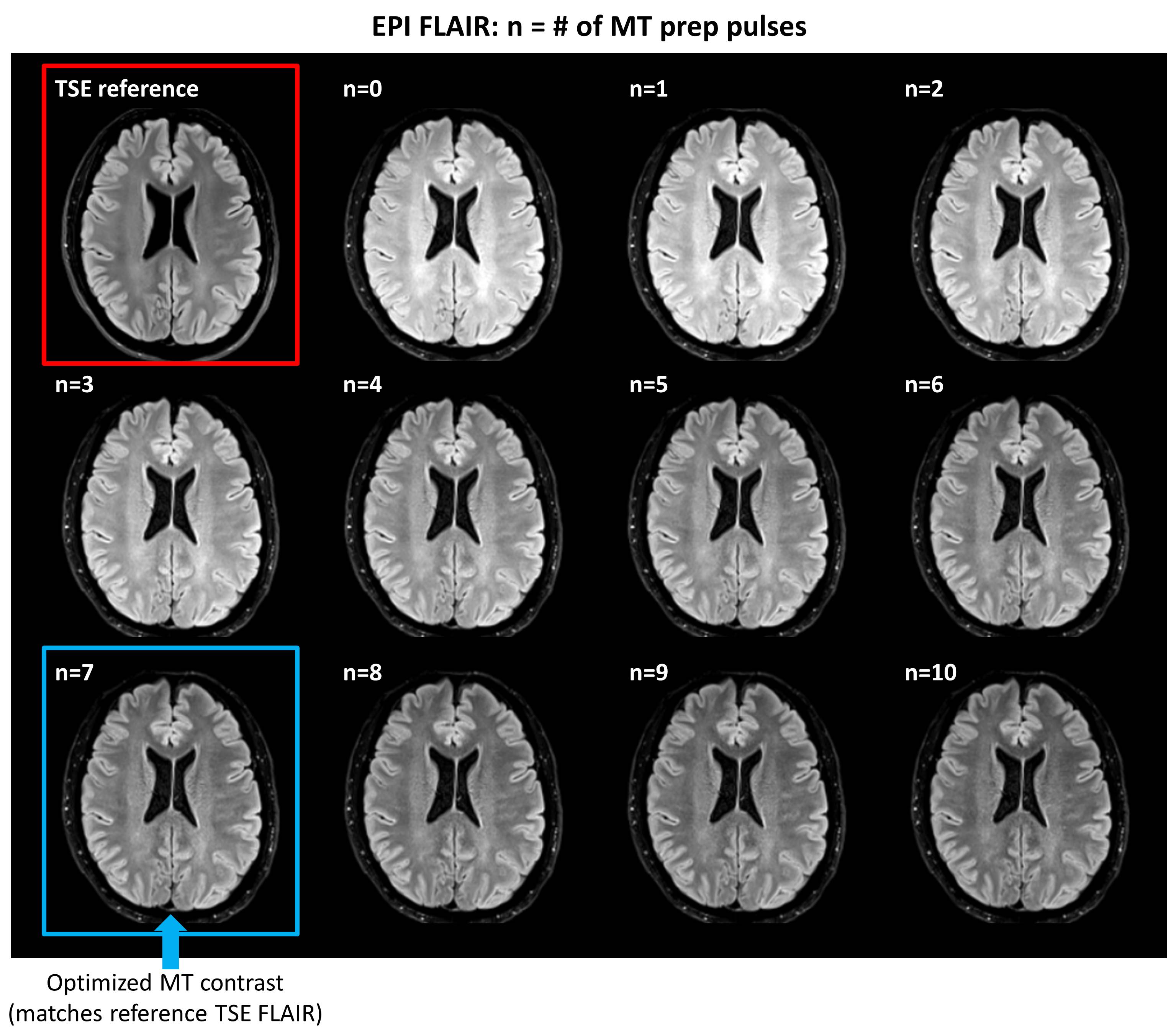
Figure 2. Representative images showing the effect of additional
magnetization preparation pulses (f=1200 Hz, a=1.0) on EPI FLAIR contrast
compared to the clinical reference TSE FLAIR scan (top left). Optimal
EPI FLAIR contrast (i.e., most closely matching the TSE reference scan) was
achieved with 7 preparation pulses (see Figure 4 for quantitative evaluation).
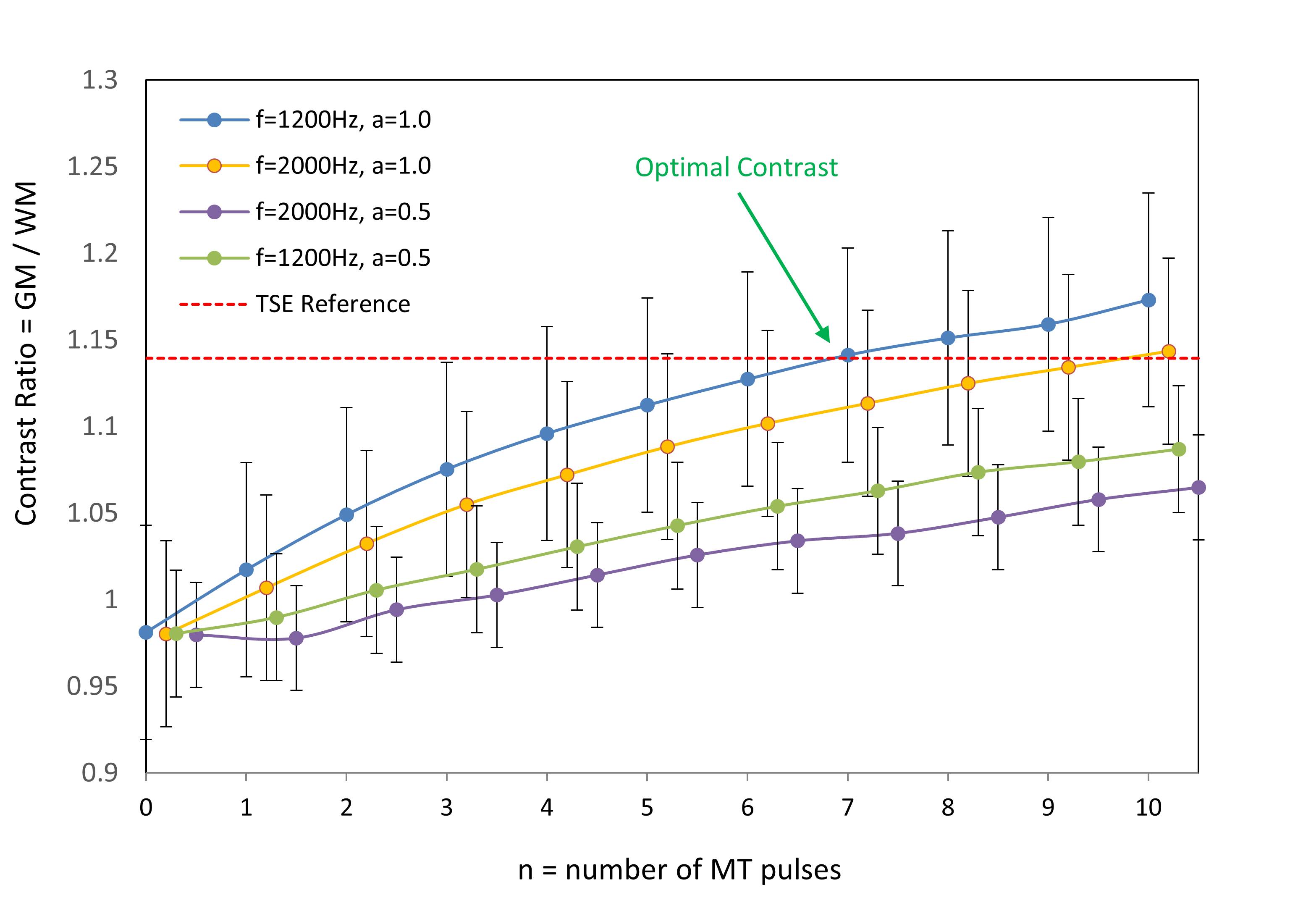
Figure 4. Contrast ratio for EPI
FLAIR images (defined as average GM / WM signal) as a function of the number of
MT preparation pulses (n), resonance offset (f), and pulse amplitude (a).
Contrast ratio for the TSE reference scan is shown by the dashed red line.
Optimal contrast (i.e., most clostly matching the TSE reference contrast) was
achieved with n=7, f=1200Hz, and a=1.0.