Alecio F. Lombardi1,2, Zhao Wei1, Hyungseok Jang1, Saeed Jerban1, Lillian Gong1, Jiang Du1, Eric Y. Chang1,2, and Ya-Jun Ma1
1Radiology, University of California, San Diego, CA, United States, 2Radiology Service, Veterans Affairs, San Diego, CA, United States
1Radiology, University of California, San Diego, CA, United States, 2Radiology Service, Veterans Affairs, San Diego, CA, United States
The 3D-DIR-UTE-Cones sequence can produce high-resolution and high-contrast imaging of the OCJ region
of the knee in vivo and shows more efficient subchondral bone marrow fat suppression compared to the IR-FS-UTE Cones sequence.
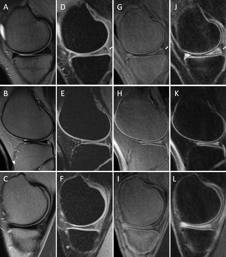
Figure 3. T2w-FSE (A, B, C), FSPGR (D, E, F), 3D IR-FS-UTE
Cones (G, H, I), and 3D DIR-UTE-Cones (J, L, M) performed in knees of two healthy
volunteers (the first row represent the first volunteer). On T2w-FSE and FSPGR,
the signal in the OCJ region cannot be detected due to its fast signal decay. (G-I)
3D IR-FS-UTE Cones sequences highlighting the OCJ and suppressing SC and
subchondral bone. (J-L) 3D DIR-UTE-Cones sequences also highlight the OCJ but
with better subchondral bone fat suppression.
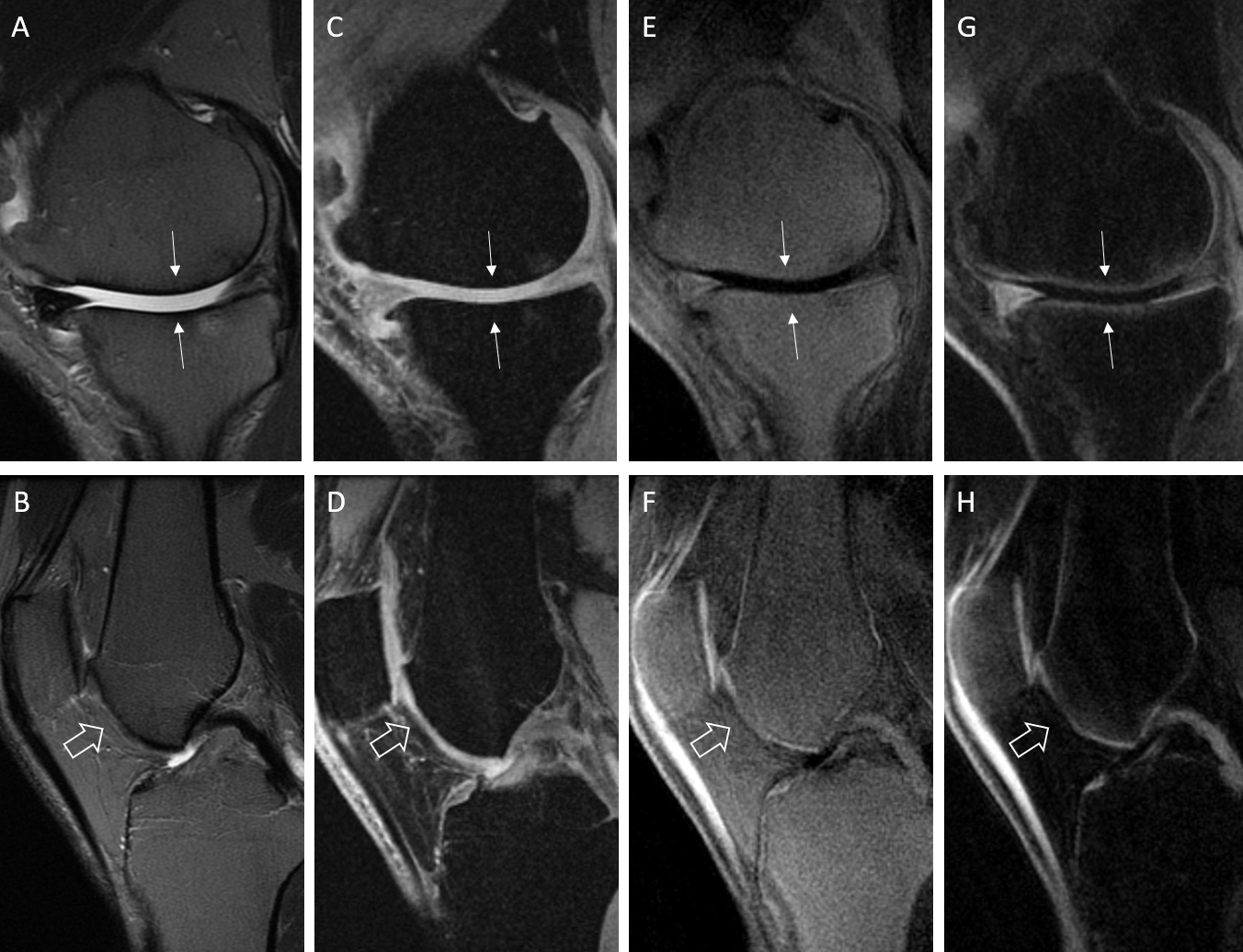
Figure 4. T2w-FSE (A, B), FSPGR (C, D), 3D IR-FS-UTE Cones
(E, F), and 3D DIR-UTE-Cones (G, H) performed in the knees of two patients with
osteoarthritis. (Arrows in A, C, E, G) show interruption of the bright-line
representing OCJ on the weight-bearing surface of the medial femoral condyle
and medial tibial plateau in the first patient and on the femoral trochlea in
the second patient (open arrows in B, D, F, H). The 3D IR-UTE-FS Cones
sequences (G and H) are more efficient in suppressing the subchondral bone fat
than the 3D IR-FS-UTE Cones (E, F).