Jinil Park1 and Jang-Yeon Park2,3
1Biomedical Institute for Convergence at SKKU, Sungkyunkwan University, Suwon, Korea, Republic of, 2Department of Biomedical engineering, Sungkyunkwan University, Suwon, Korea, Republic of, 3Department of Intelligent Precision Healthcare Convergence, Sungkyunkwan University, Suwon, Korea, Republic of
1Biomedical Institute for Convergence at SKKU, Sungkyunkwan University, Suwon, Korea, Republic of, 2Department of Biomedical engineering, Sungkyunkwan University, Suwon, Korea, Republic of, 3Department of Intelligent Precision Healthcare Convergence, Sungkyunkwan University, Suwon, Korea, Republic of
this study demonstrates VS-UTE ability to maintain image quality when the number of projection views is undersampled or when the imaging volume is selected smaller than the imaging target.
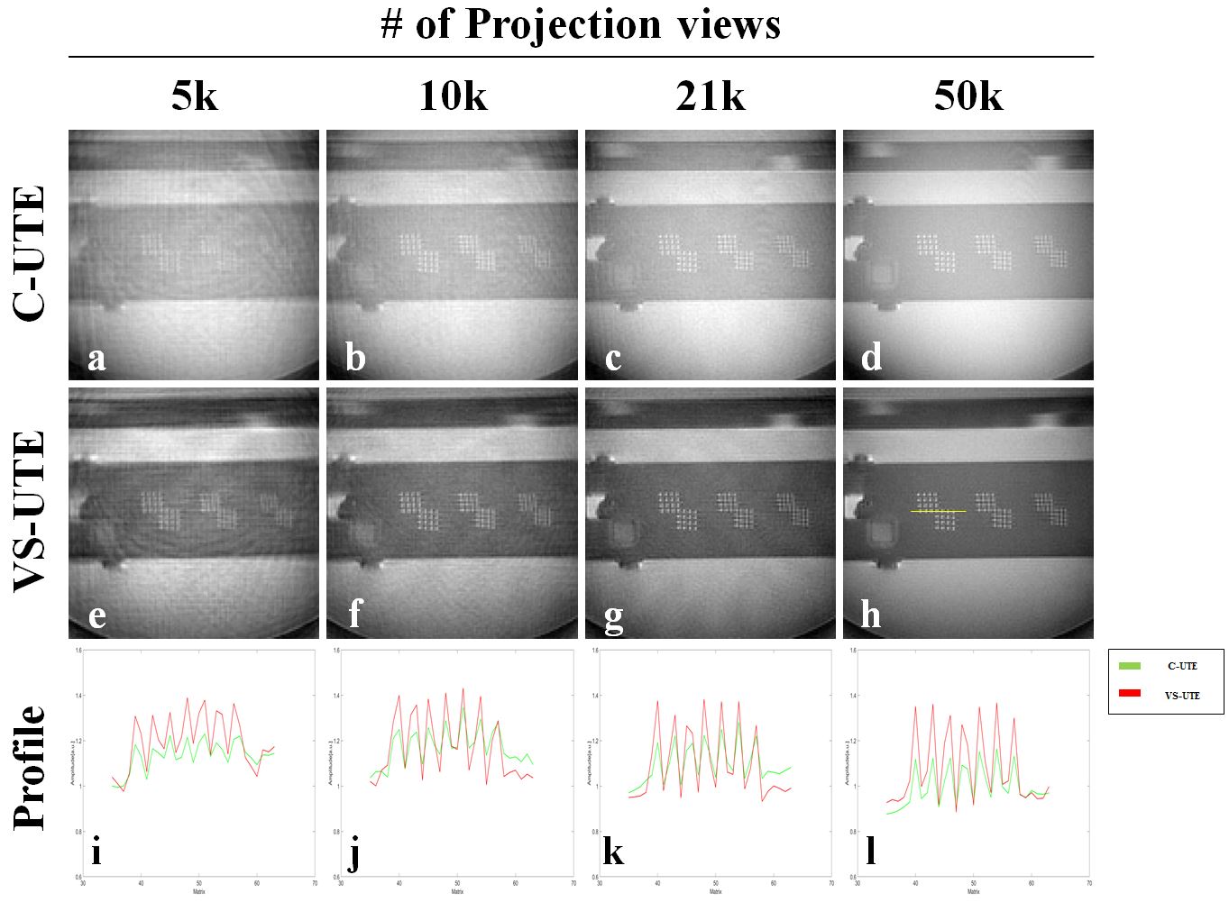
Figure 3. the images including the resolution grids and 1D profiles of the ACR phantom acquired
using conventional UTE (C-UTE) (a-d) and VS-UTE (e-h). The C-UTE image is
difficult to show the internal structure due to the phantom signal outside the
ROI when the number of projections was 5k, but VS-UTE provided a better internal structure image. Also, the
overall CNR (contrast to noise ratio) of C-UTE was lower than that of
VS-UTE(i-l).
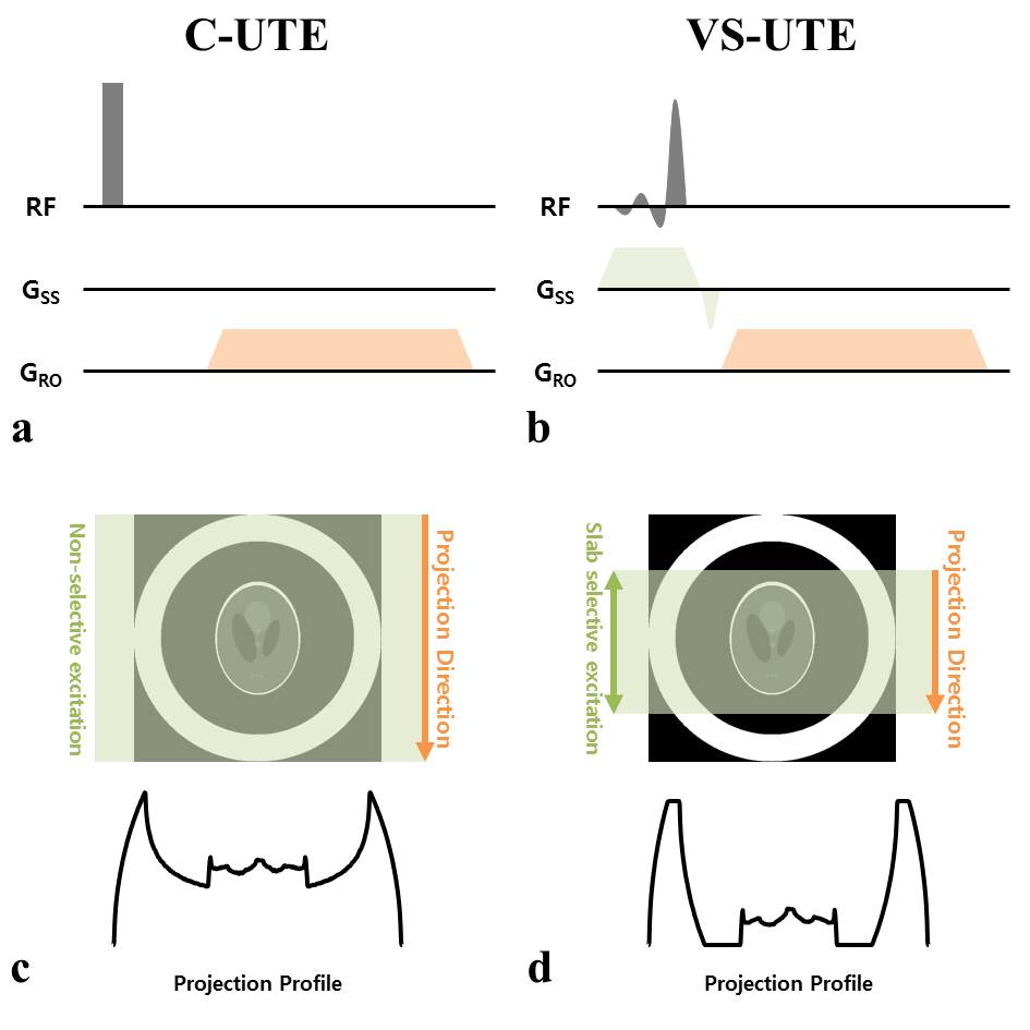
Figure 1. Pulse sequence diagrams(a, b)
and data acquisition method(c, d) of conventional UTE (C-UTE, a) and
volume-selective UTE (VS-UTE, b). C-UTE, which uses a non-selective pulse for
spin excitation(a), acquires projection data including spin information of the
entire object(c). In contrast, The spin excitation of the unexcited object is
not included in the signal(d) by using the slab selective pulse and the slab
selective gradient simultaneously and setting the projection direction in the
same direction as the slab selection direction(b).