Rolf F Schulte1, Ana Beatriz Solana1, and Scott R Hinks2
1GE Healthcare, Munich, Germany, 2GE Healthcare, Waukesha, WI, United States
1GE Healthcare, Munich, Germany, 2GE Healthcare, Waukesha, WI, United States
- EPI and Spiral imaging were with combined with BURST, where magnetisation is excited with a train of RF pulses
- Off-resonance artefacts are reduced by splitting the long single-shot readout trajectories into multiple segments acquired inside the same pulse-and-acquire block
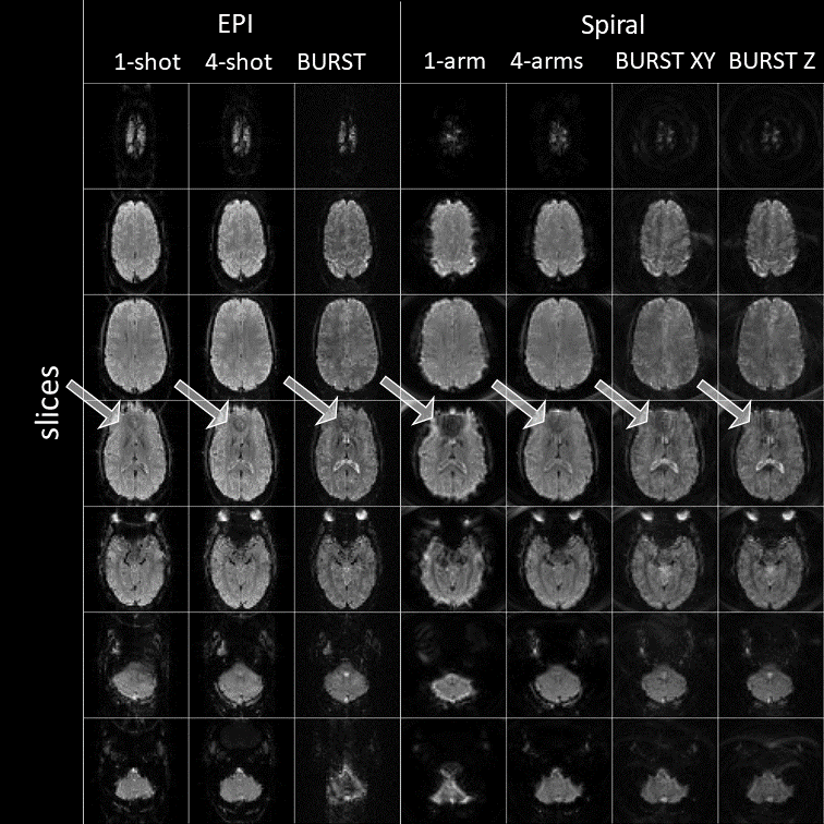
Fig. 3: Sequence comparison results in the brain of a healthy volunteer. A
particularly sensitive area are the sinuses (depicted by arrows). B0
inhomogeneities again lead to geometric distortions (EPI), blurring (Spirals)
and also signal dropout, particularly in case of the single shot spiral.
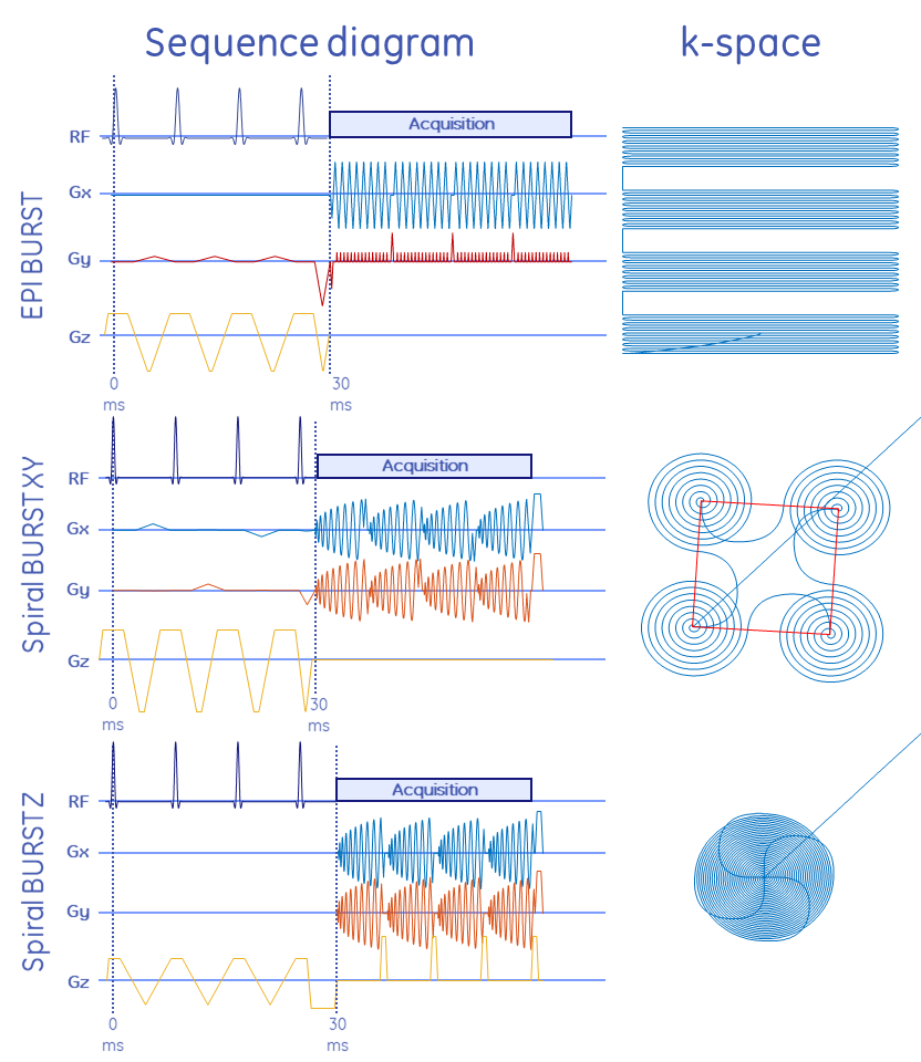
Fig. 1: Sequence diagram and corresponding k-space of
the 3 BURST sampling schemes. A train of 4 slice-selective RF pulses excites
magnetisation, storing it in outer parts of k-space through gradients in
(logical) xy (top and middle) or z direction (bottom). The readout is split
into 4 segments, and each respective sub-RF pulse is refocused in centre of
k-space (central line of EPI and beginning of Spiral trajectory). Hence, in the
k-space depiction, the 4 separate regions in k-space all encode a similar area
in k-space with EPI shifted by a quarter of a line and Spiral interleaved by
90°.