Jin Jin1,2, Monique Tourell2, Pascal Sati3, Sunil Patil4, Kecheng Liu5, John Derbyshire6, Fei Han7, Saskia Bollmann2, Simon Robinson2, Josef Pfeuffer8, Steffen Bollmann2, Markus Barth2, and Kieran O'Brien1
1Siemens Healthcare, Brisbane, Australia, 2The University of Queensland, Brisbane, Australia, 3Cedars-Sinai Medical Center, Los Angeles, CA, United States, 4Siemens Medical Solutions USA, Baltimore, MD, United States, 5Siemens Medical Solutions USA, Baltimore, OH, United States, 6National Institute of Mental Health, Bethesda, MD, United States, 7Siemens Medical Solutions USA, Los Angeles, CA, United States, 8Siemens Healthcare, Sydney, Australia
1Siemens Healthcare, Brisbane, Australia, 2The University of Queensland, Brisbane, Australia, 3Cedars-Sinai Medical Center, Los Angeles, CA, United States, 4Siemens Medical Solutions USA, Baltimore, MD, United States, 5Siemens Medical Solutions USA, Baltimore, OH, United States, 6National Institute of Mental Health, Bethesda, MD, United States, 7Siemens Medical Solutions USA, Los Angeles, CA, United States, 8Siemens Healthcare, Sydney, Australia
A 3D EPI provides
a flexible combination of single/multi shot scheme, in-plane
segmentation, echo-train length, partial Fourier, and 2D-GRAPPA/2D-CAIPIRINHA acceleration. Highly accelerated high-resolution
susceptibility-based imaging with markedly reduced scan time is presented.
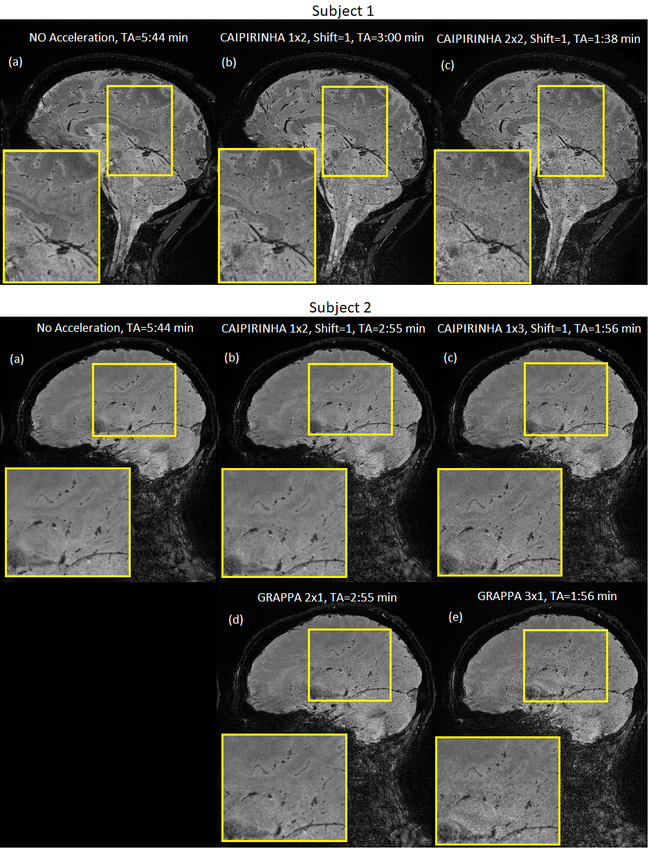
Figure 4 T2*-weighted 3D EPI images acquired using the proposed implementation
with 2 subjects. Imaging
protocols are summarized shown in Figure 3. Sections of the images were
enlarged for detailed examination.
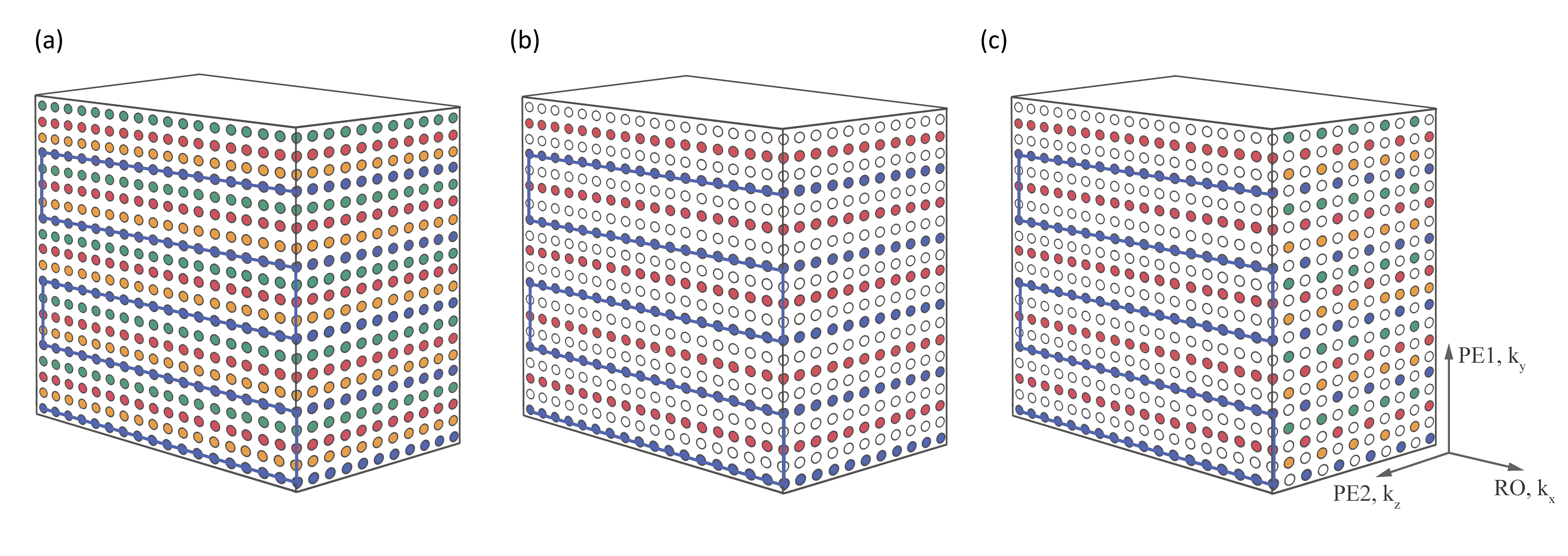
Figure 1 Examples of k-Space sampling patterns when fully-sampled (a), and with
GRAPPA 2×1 (b) and CAIPIRINHA 1×2 (c) accelerations. (a): The 3D EPI volume was
separated into shots in the kx-ky plane (four shots in this case; blue, yellow,
red and green) and partitions in the kz direction. (b): The GRAPPA acceleration
was achieved by skipping the 2nd and 4th shots for all
the partitions. (c): The CAIPIRINHA acceleration was achieved by alternatively
skipping 2nd and 4th shots and then 1st and 3rd
shots between successive partitions.