Kaibao Sun1, Zheng Zhong1,2, Guangyu Dan1,2, Muge Karaman1,2, Qingfei Luo1, and Xiaohong Joe Zhou1,2,3
1Center for MR Research, University of Illinois at Chicago, Chicago, IL, United States, 2Department of Bioengineering, University of Illinois at Chicago, Chicago, IL, United States, 3Departments of Radiology and Neurosurgery, University of Illinois at Chicago, Chicago, IL, United States
1Center for MR Research, University of Illinois at Chicago, Chicago, IL, United States, 2Department of Bioengineering, University of Illinois at Chicago, Chicago, IL, United States, 3Departments of Radiology and Neurosurgery, University of Illinois at Chicago, Chicago, IL, United States
A 3D reduced
field-of-view imaging (3D-rFOVI) sequence based on a slab-selective 2D RF pulse
has produced images with high isotropic spatial resolution and reduced image
distortion that are useful in fMRI and DWI.
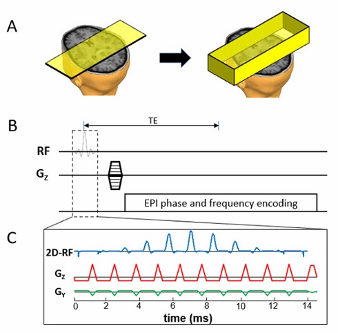
Figure 1: (A): A schematic to
illustrate 3D rFOVI by replacing slice selection with slab selection using a 2D
RF pulse in (C), followed by through-slab phase-encoding as shown in (B). (B): A
conceptual 3D rFOVI sequence with phase-encoding gradient along the slab
direction. (C): Details of the 2D RF excitation pulse with a fly-back EPI excitation
k-space trajectory to avoid the issues associated with Nyquist ghosts.
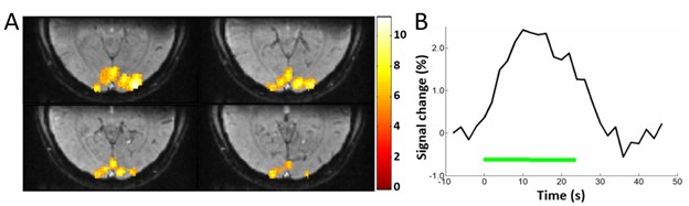
Figure 4: (A): Visual fMRI activation maps, selected
from the 3D volume, overlaid onto the baseline images acquired with 3D-rFOVI GRE-EPI.
(B): The averaged time course showing the activation. The signal change in (B)
is approximately 2.5%. The green bar represents the time period for visual
stimulus, which was 24s.