Jessica Schäper1,2, Grzegorz Bauman1,2, Carl Ganter3, and Oliver Bieri1,2
1Department of Biomedical Engineering, University of Basel, Basel, Switzerland, 2Department of Radiology, Division of Radiological Physics, University Hospital Basel, Basel, Switzerland, 3Department of Diagnostic and Interventional Radiology, Klinikum rechts der Isar, Technical University of Munich, Munich, Germany
1Department of Biomedical Engineering, University of Basel, Basel, Switzerland, 2Department of Radiology, Division of Radiological Physics, University Hospital Basel, Basel, Switzerland, 3Department of Diagnostic and Interventional Radiology, Klinikum rechts der Isar, Technical University of Munich, Munich, Germany
For some tissues the frequency response of bSSFP exhibits a pronounced asymmetry due to the intra-voxel frequency distributions. In this work we show that this asymmetry disappears for brain tissue in the limit of $$$\mathit{TR}\sim 1\,\mathrm{ms}$$$ at $$$3\,\mathrm{T}$$$.
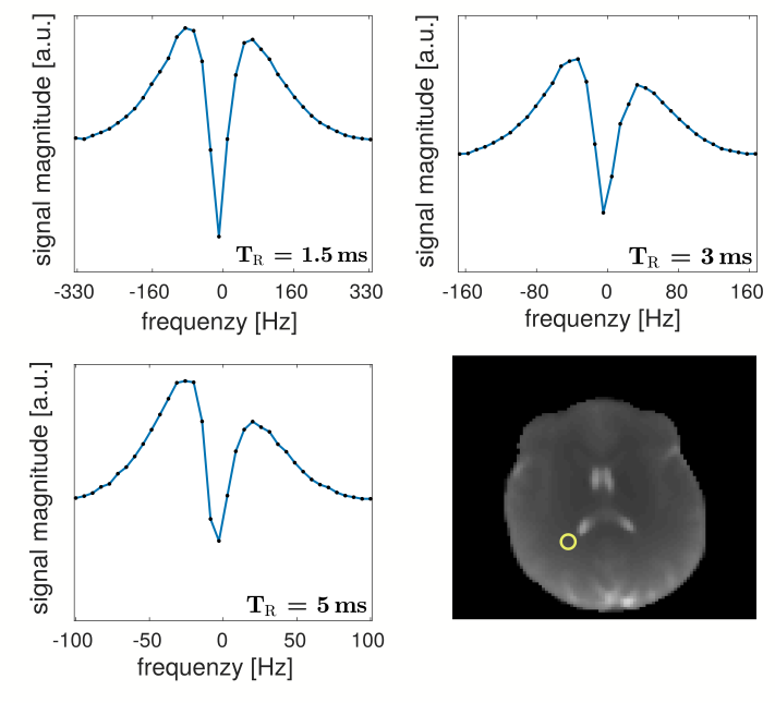
The bSSFP profiles for the mean value of a white matter ROI, shown in the reference on the bottom right, are displayed. The asymmetry for $$$\mathit{TR}=5\,\mathrm{ms}$$$ $$$(\mathit{AI}=0.14)$$$ and $$$\mathit{TR}=3\,\mathrm{ms}$$$ $$$(\mathit{AI}=0.08)$$$ is clearly visible, while the profile for $$$\mathit{TR}=1.5\,\mathrm{ms}$$$ $$$(\mathit{AI}=0.02)$$$ exhibits almost no asymmetry.
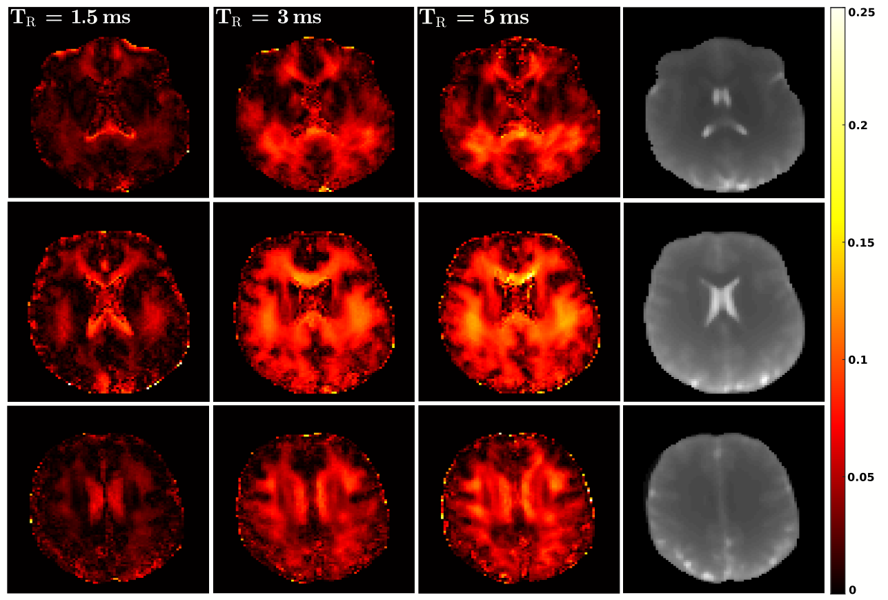
The calculated $$$\mathit{AI}$$$ maps for the different $$$\mathit{TR}$$$ are shown for three different axial slices of the in vivo brain scans. It is clearly visible that, overall, the values for $$$\mathit{TR} = 1.5\,\mathrm{ms}$$$ are smaller than for $$$\mathit{TR} = 3\,\mathrm{ms}$$$ and $$$\mathit{TR} = 5\,\mathrm{ms}$$$. The right pictures show anatomical references.