Matthijs de Buck 1, Aaron Hess2, and Peter Jezzard1
1Wellcome Centre for Integrative Neuroimaging, FMRIB Division, Nuffield Department of Clinical Neurosciences, University of Oxford, Oxford, United Kingdom, 2Oxford Centre for Clinical Magnetic Resonance Research, Department of Cardiovascular Medicine, University of Oxford, Oxford, United Kingdom
1Wellcome Centre for Integrative Neuroimaging, FMRIB Division, Nuffield Department of Clinical Neurosciences, University of Oxford, Oxford, United Kingdom, 2Oxford Centre for Clinical Magnetic Resonance Research, Department of Cardiovascular Medicine, University of Oxford, Oxford, United Kingdom
Optimized DANTE-preparation
when using DANTE-SPACE at 7T increases the vessel wall visibility while
reducing the SAR.
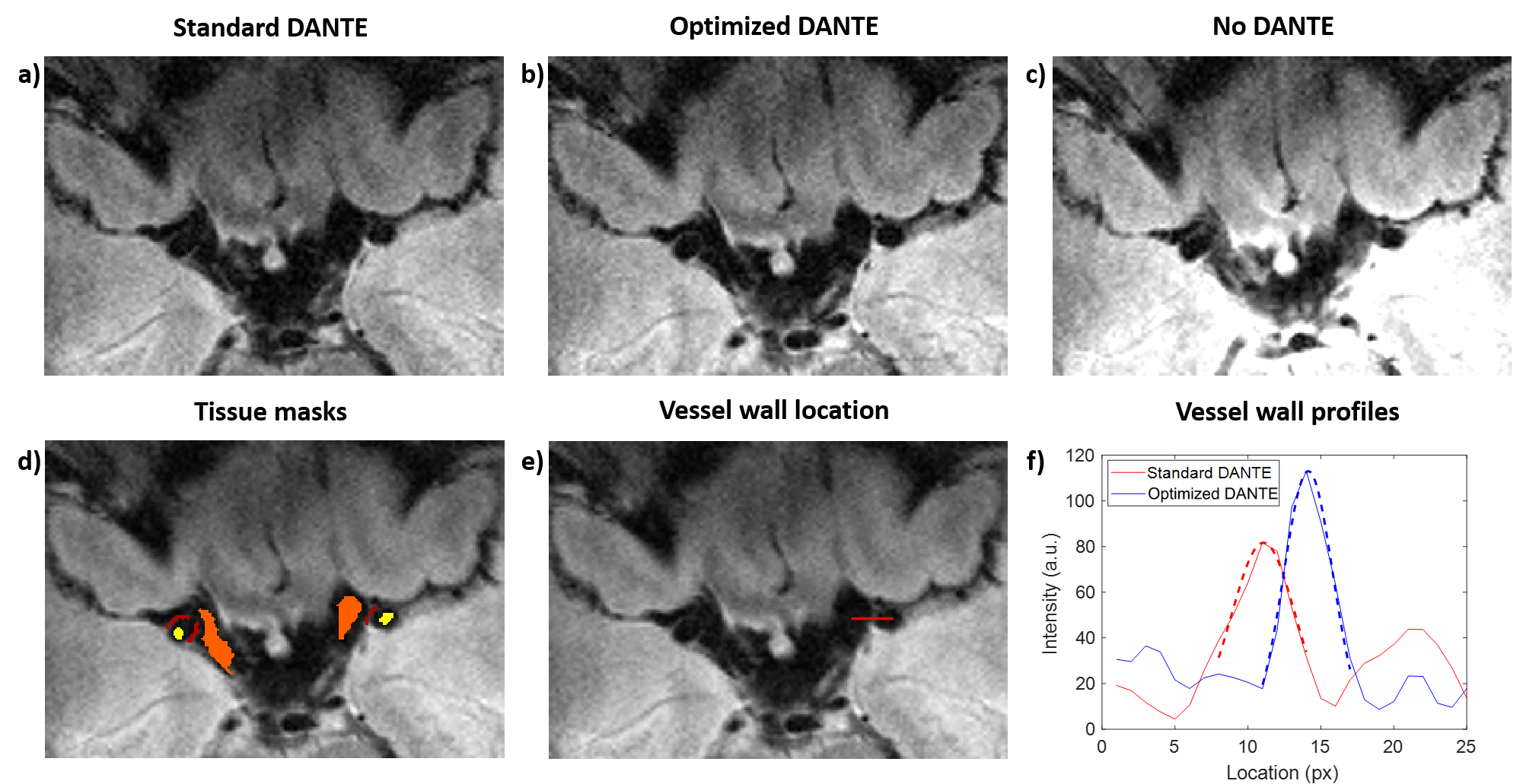
Figure 4: DANTE-SPACE data acquired using (a) 300 DANTE-pulses of 10 degrees, (b)
200 DANTE-pulses of 9 degrees, and (c) without DANTE-preparation. (d-e)
The mean of Figures (a) and (b), showing (d) conservatively drawn tissue
masks for the vessel wall (red), CSF (orange), and lumen (yellow), and (e)
an example of a vessel wall location (red line). (f) The corresponding
vessel wall profiles using “standard” DANTE-preparation (a) and “optimized” DANTE-preparation
(b). Dashed lines indicate Gaussian fits to the 7 pixels surrounding the peaks.
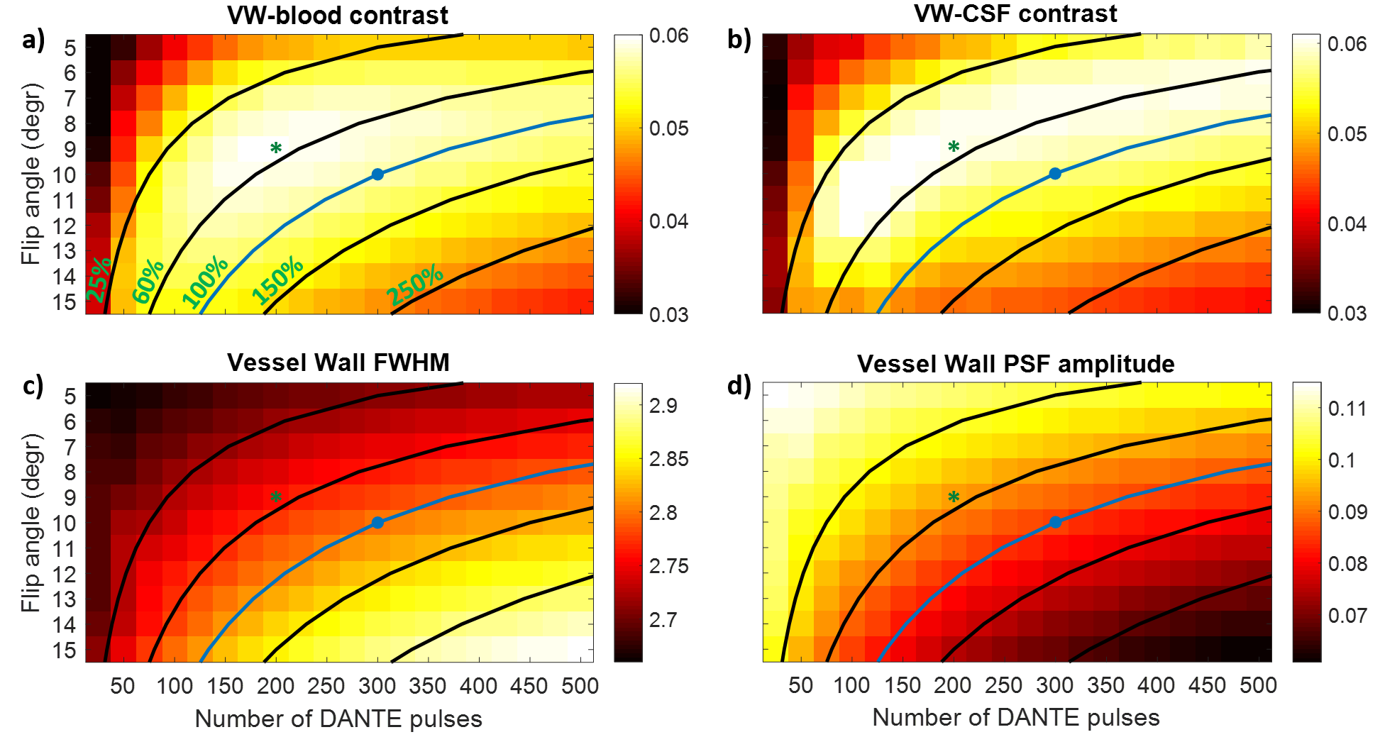
Figure 3: Simulations of DANTE-SPACE using different DANTE-parameters. Blue
dots correspond to “standard” DANTE; green asterisks to “optimized” DANTE. Black
and blue lines indicate iso-contour SAR-lines of the DANTE-preparation (which
accounts for approximately 30% of the total SAR in DANTE-SPACE), relative to the SAR of
“standard” DANTE; values for each line are shown in (a). Figures show (a)
contrast between the vessel wall and blood, (b) contrast between the
vessel wall and CSF, (c) the FWHM of the vessel wall signal (px.), and (d)
intensity of the vessel wall signal.