Yayun Ji1,2, Jianqiang Fang2, Binyu Zhang2, Siyuan Mi2, Weiyin Vivian Liu3, Weian Zhao2, and Liheng Ma1
1The First Affiliated Hospital of Guangdong Pharmaceutical University, Guangzhou, China, 2Xianyang Central Hospital, Xianyang, China, 3MR research, GE Healthcare, Beijing, China
1The First Affiliated Hospital of Guangdong Pharmaceutical University, Guangzhou, China, 2Xianyang Central Hospital, Xianyang, China, 3MR research, GE Healthcare, Beijing, China
This study aims to investigate if advanced UTE T2* Mapping MRI quantitative sequence could sensitively assess the early changes in the biochemical components of the cartilage endplate.
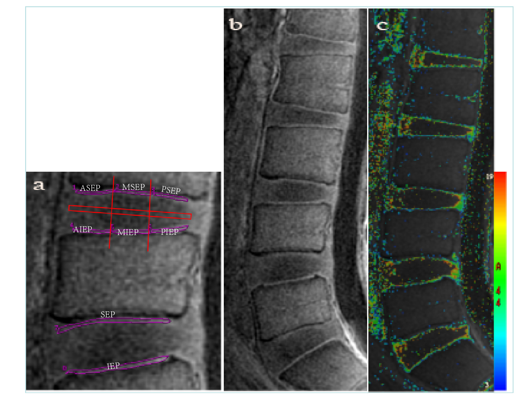
Figure 1 UTE T2*
Mapping of cartilaginous endplate. (a)
the cartilage endplates of L1/2-L5/S1 were divided into three equal parts from
front to back, and the ROI of each part was delineated, including ASEP, MSEP,
PSEP, AIEP, MIEP and PIEP. Then, the
superior and inferior cartilaginous endplates (SEP, IEP) were completely
delineated, that is, the complete measurement was carried out without
triserification, and then the T2* value of each part was measured in false
color figure b and c
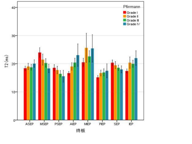
FIG. 2 Bar plots of T2* in the Pfirrmann graded
cartilaginous endplate in four groups. Except
for Grade I, the T2* value of Grade III-V in the superior endplate was inferior
than that in the inferior endplate. T2* value of superior endplate decreases
gradually from Grade I-IV. The value of T2* in the inferior and rear end of the
superior endplate was inferior than that in the front middle. T2* in the
posterior part of the superior endplate was higher than that in the inferior
end plate for Grade I-II and opposite for Grade III-IV.