Cui Ren1, Qing Li2, Stefan Sommer3,4, Qiao Zhu1, and Huishu Yuan1
1Radiology, Peking University Third Hospital, Beijing, China, 2MR Collaborations, Siemens Healthcare Ltd, Shanghai, China, 3Siemens Healthcare, Zurich, Switzerland, 4Swiss Center for Musculoskeletal Imaging (SCMI), Zurich, Switzerland
1Radiology, Peking University Third Hospital, Beijing, China, 2MR Collaborations, Siemens Healthcare Ltd, Shanghai, China, 3Siemens Healthcare, Zurich, Switzerland, 4Swiss Center for Musculoskeletal Imaging (SCMI), Zurich, Switzerland
Compared with conventional imaging of the sacroiliac joint for ankylosing spondylitis (AS) assessment, 3D-UTE provides quantitative T2* values. These are significantly higher in AS compared with controls, providing potential diagnostic value.
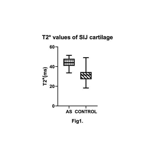
Fig1. Average T2* values of the AS group were statistically higher than those of the control group.
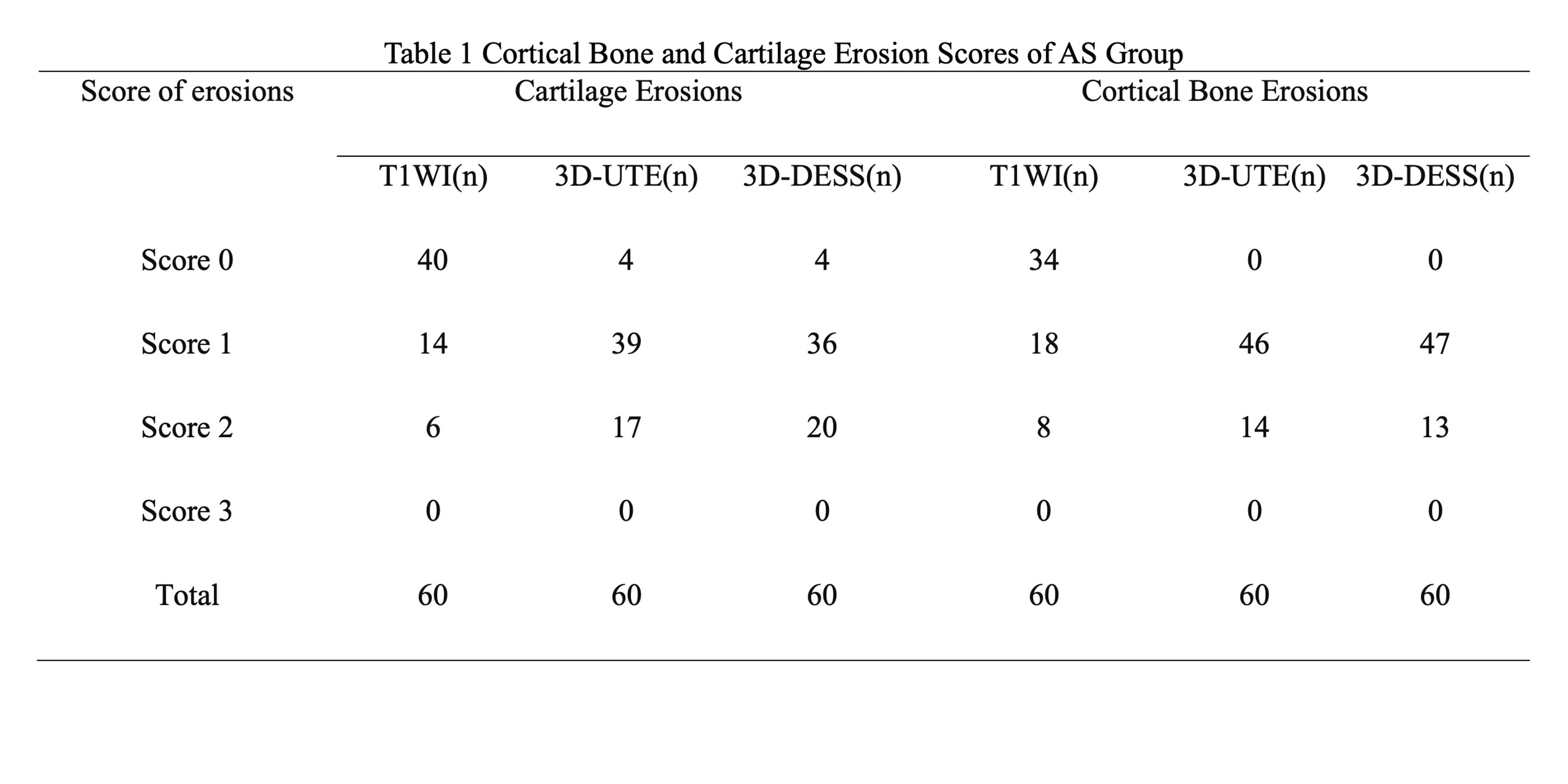
Note. — A total of 30 AS patients with 60 sacroiliac joints; n, numbers of sacroiliac joints