Victor Babu Kassey1,2,3,4, Matthias Walle1, Jonathan Egan1, Diana Yeritsyan1, Yaotang Wu2,3,4, Brian D Snyder2,4, Edward Rodriguez1,2, Jerome Ackerman2,5, and Ara Nazarian1,2,4
1Carl J. Shapiro Department of Orthopaedic Surgery, Beth Israel Deaconess Medical Center, Boston, MA, United States, 2Harvard Medical School, Boston, MA, United States, 3Massachusetts General Hospital, Charlestown, MA, United States, 4Department of Orthopaedic Surgery, Boston Children's Hospital, Boston, MA, United States, 5Department of Radiology, Massachusetts General Hospital, Charlestown, MA, United States
1Carl J. Shapiro Department of Orthopaedic Surgery, Beth Israel Deaconess Medical Center, Boston, MA, United States, 2Harvard Medical School, Boston, MA, United States, 3Massachusetts General Hospital, Charlestown, MA, United States, 4Department of Orthopaedic Surgery, Boston Children's Hospital, Boston, MA, United States, 5Department of Radiology, Massachusetts General Hospital, Charlestown, MA, United States
Solid-state 1H
and 31P ZTE imaging were feasible to compute bone matrix and mineral
densities on ex-vivo rat bone specimens at 7T. We expect that this
method can be extended to humans to measure bone matrix and mineral densities to
identify osteoporosis and osteomalacia non-invasively.
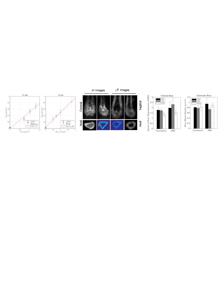
Figure 1(a): The least
squares fit of known PEG and HA phantom densities vs. 1H and 31P MRI
derived intensities after B1 correction
with 3-point calibration. (b). Rat femur 1H images (water+fat suppressed)
and 31P
images, the same slice 1H & 31P images (blue color). (c). EBM values from
Gravimetry and MRI analysis.