Yijie Fang1, Wenjun Yu1, Long Qian2, and Shaolin Li1
1The Fifth Affiliated Hospital of Sun Yat-sen University, Zhuhai, China, 2MR Research, GE Healthcare, Beijing, China
1The Fifth Affiliated Hospital of Sun Yat-sen University, Zhuhai, China, 2MR Research, GE Healthcare, Beijing, China
Synthetic MRI technique has a good display of knee joint structural lesions, and is of high value in monitoring the dynamic changes of knee cartilage extracellular matrix in amateur marathon athletes
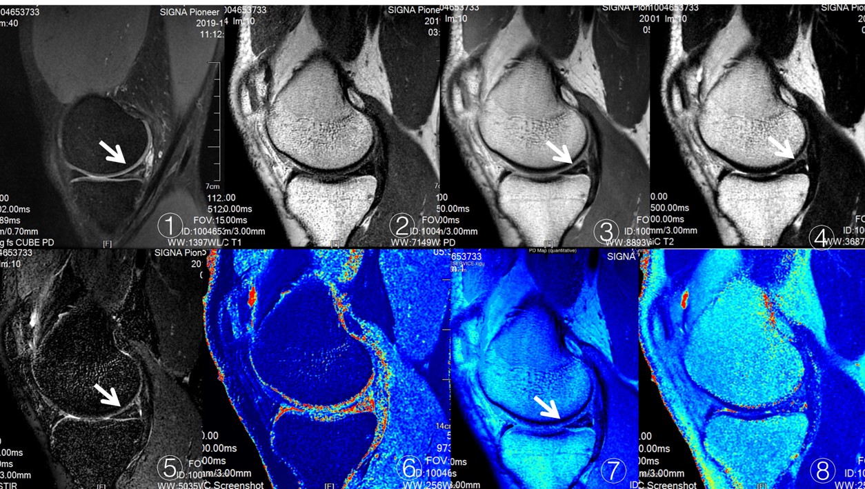
Male, 35 years old, participated in the full marathon twice, and his medial half month later his foot was torn horizontally. Fig. ① - ⑧ showed 3d Cube-Pd and Magic (T1WI, IW, T2WI, stir, t1mapping, pdmapping, T2mapping) respectively. ① 3D cube-pd showed linear hyperintensity in the posterior horn of the medial meniscus, magiciw, T2WI and stir sequences showed mild hyperintensity in the posterior horn of the meniscus. figure ⑦ Pd-mapping. The signal intensity of medial meniscus was slightly increased in T1 mapping and figure ⑧ in T2WI mapping.
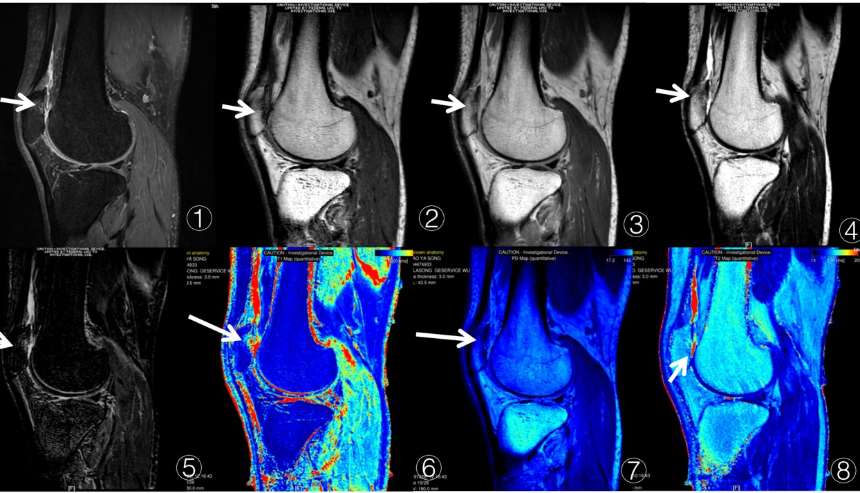
Male, 45 year old, participated in the whole marathon for 5 times, suffered from patellar cartilage injury. Fig. ① 3D cube-pd shows the absence of the inferior part of patellar cartilage, bone marrow edema under the articular surface, magec T1WI sequence, IW and T2WI show the absence of cartilage, and the bone marrow edema under the articular surface is not clear. Figure ⑦ PD mapping showed partial cartilage defect and slight increase of bone marrow edema signal. T2WI mapping showed that the signal intensity in some areas increased, but no obvious signal increase in bone marrow edema