Hanqi Wang1, Qing Li2, Stefan Sommer3,4, and Yong Lu1
1Ruijin Hospital, School of Medicine, Shanghai Jiao Tong University, Shanghai, China, 2MR Collaboration, Siemens Healthcare Ltd., Shanghai, China, Shanghai, China, 3Siemens Healthcare, Zurich, Switzerland, Zurich, Switzerland, 4Swiss Center for Musculoskeletal Imaging (SCMI), Balgrist Campus, Zurich, Switzerland, Zurich, Switzerland
1Ruijin Hospital, School of Medicine, Shanghai Jiao Tong University, Shanghai, China, 2MR Collaboration, Siemens Healthcare Ltd., Shanghai, China, Shanghai, China, 3Siemens Healthcare, Zurich, Switzerland, Zurich, Switzerland, 4Swiss Center for Musculoskeletal Imaging (SCMI), Balgrist Campus, Zurich, Switzerland, Zurich, Switzerland
Quantitative MRI using UTE is used to show the differences in mechanical loading between deep and superficial cartilage, which is reflective of different bio-mechanical properties in these tissues.
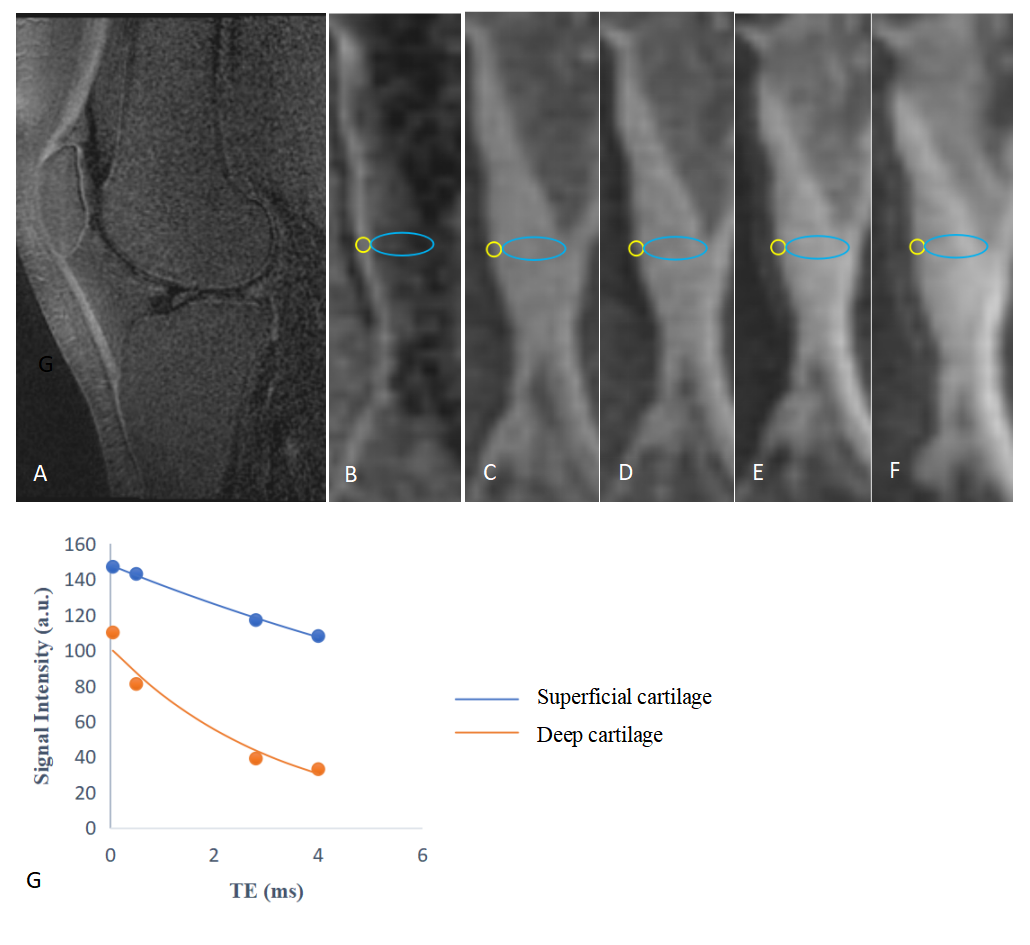
Figure 1. Example UTE subtraction images of patella cartilage (A, B, TE1 = 0.1 ms, TE2 = 2.8 ms) , as well as UTE images at a series of TEs: (C) 0.1 ms, (D) 0.5 ms, (E) 2.8 ms, and (F) 5 ms. ROIs were delineated on deep (yellow) and superficial (blue) layers of patella cartilage in B and copied to C-F to measure the signal intensity. T2* values from single-component exponential fitting curves (G) were 3.08 ms deep patella cartilage and 13.28 ms superficial patella cartilage.
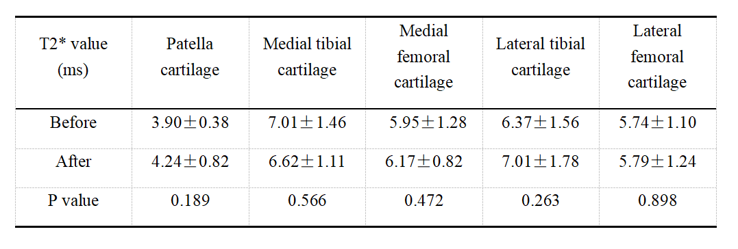
Table 1. Deep cartilage T2* values before and after activity