1Radiology, University Hospital Tuebingen, Germany, Tuebingen, Germany, 2Siemens Healthineers, Forchheim, Germany
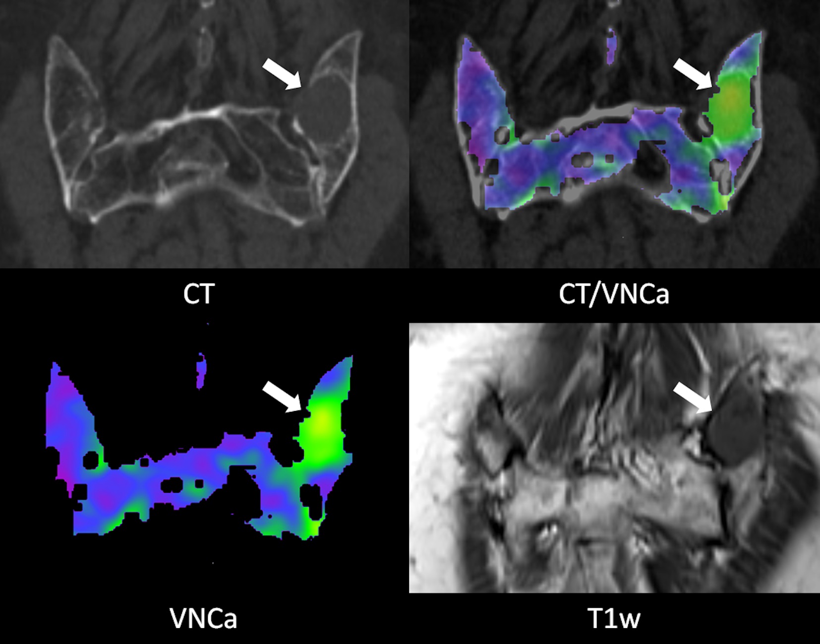
Figure 2
Example images of a 74 year old female patient with progressive multiple myeloma (first diagnosis 5 years and 3 months prior). The focal osteolytic lesion in the left dorsal iliac crest (arrow) is conspicuous in the CT as well as the VNCa image (mean VNCa attenuation 42.3 HU). In the T1w MRI image the lesion shows a homogeneous hypointense signal.
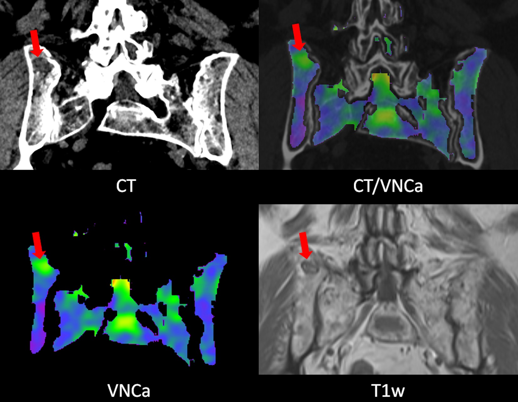
Figure 3
Example images of a 69 year old female patient with multiple myeloma in partial response (first diagnosis 3 years and 1 month prior). The focal osteolytic lesion in the right dorsal iliac crest is visible but not conspicuous in the CT image. The lesion is nicely depicted in the VNCa image (mean VNCa attenuation 7.6 HU). In the T1w MRI image the lesion shows moderate central hypointensity and a markedly hypointense rim which does not represent the typical appearance of an “active” lesion but is consistent with partial remission.