Zhao Wei1,2,3, Hyungseok Jang1, Zubiad Ibrahim1, Mohammadamin Cheraghi1, Graeme M. Bydder1, Wenhui Yang2,3, and Ya-Jun Ma1
1Department of Radiology, UC San Diego, San Diego, CA, United States, 2Institute of Electrical Engineering, Chinese Academy of Sciences, Beijing, China, 3University of Chinese Academy of Sciences, Beijing, China
1Department of Radiology, UC San Diego, San Diego, CA, United States, 2Institute of Electrical Engineering, Chinese Academy of Sciences, Beijing, China, 3University of Chinese Academy of Sciences, Beijing, China
The 3D UTE-AFI-STR method provides accurate T1
mapping of cortical bone with improved time efficiency as compared with
UTE-AFI-VTR method.
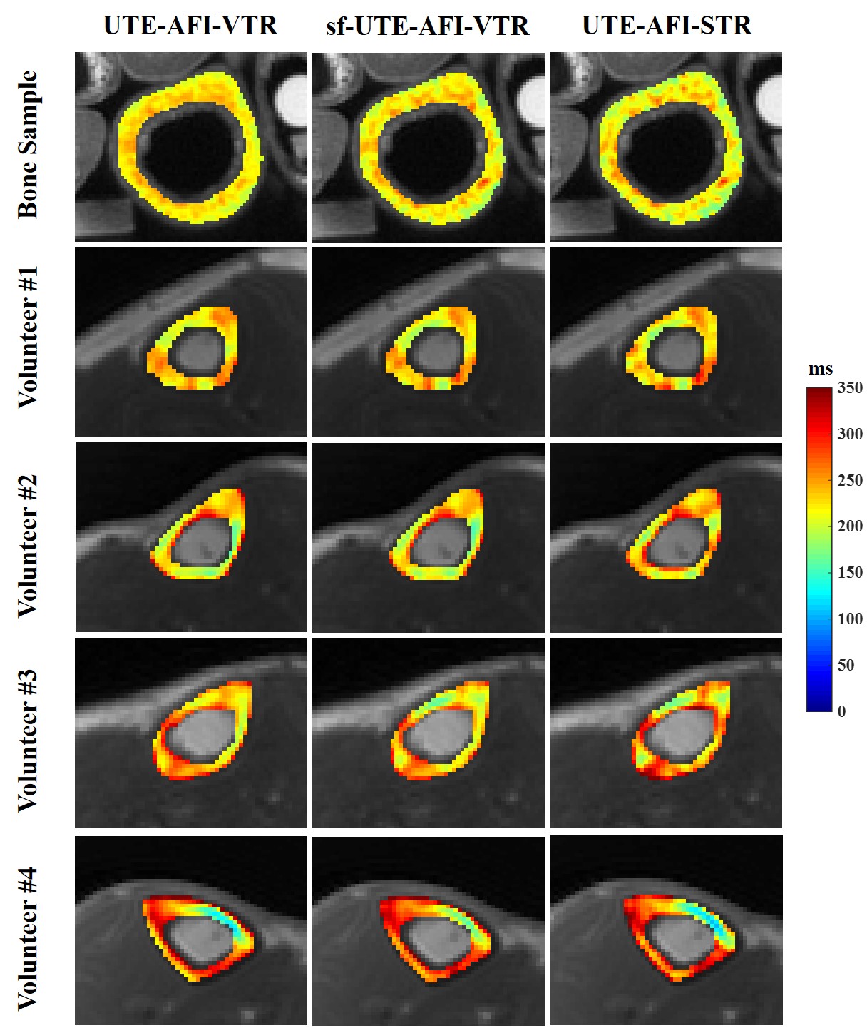
Figure
3.
Representative ex vivo human cortical bone sample (first row) and in vivo
tibial cortical bone T1 maps of four healthy volunteers (second to
fifth row) generated using the UTE-AFI-VTR (first column), the sf-UTE-AFI-VTR
(second column) and the UTE-AFI-STR (third column) methods. Similar appearances
on the T1 maps are seen with each of the three methods.
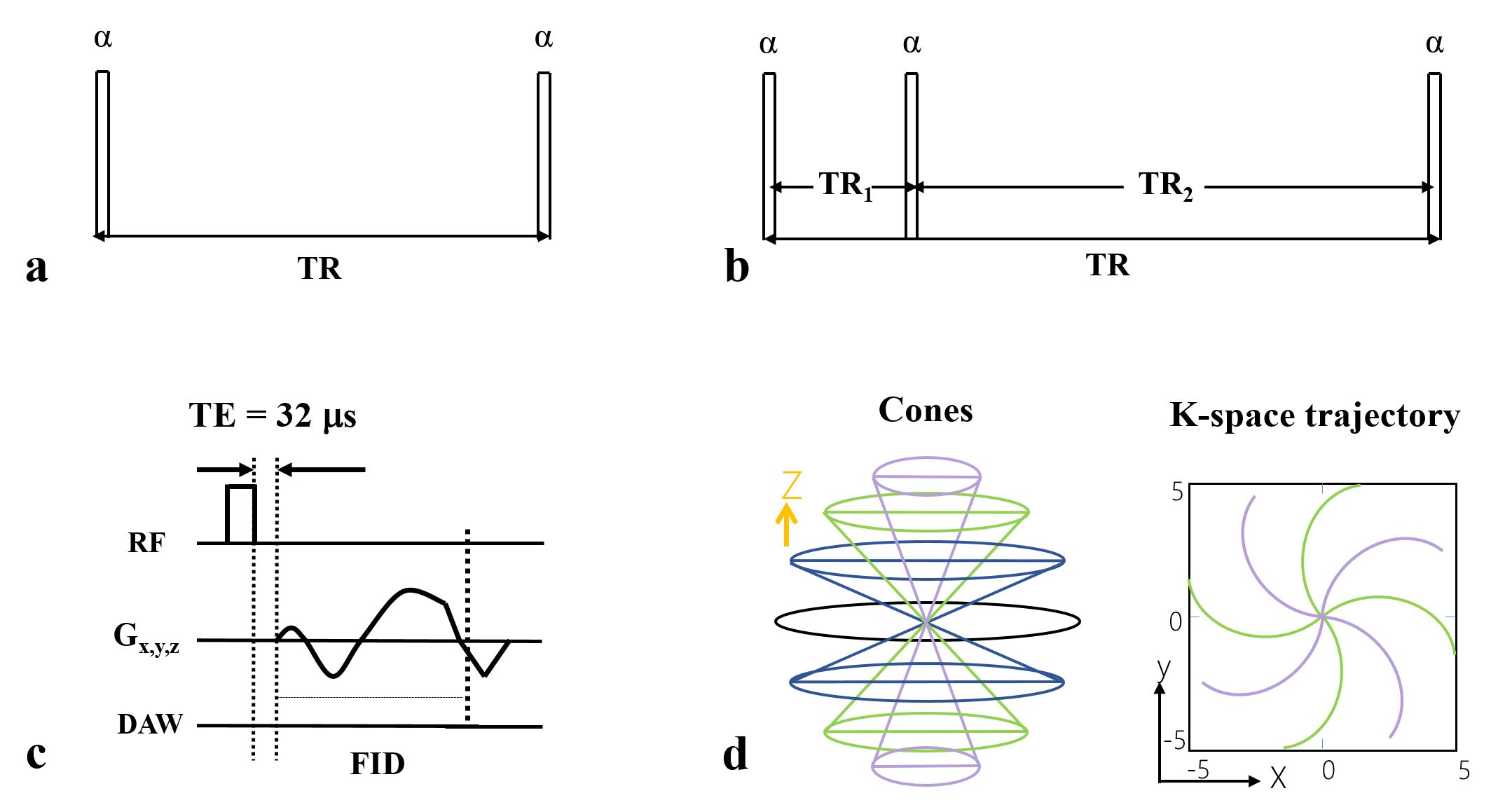
Figure 1. 3D UTE-based AFI and VTR sequences. The conventional
3D UTE sequence is used for multiple-TR data acquisition (a). The 3D UTE-AFI sequence acquires data with two interleaved TRs
(TR1 and TR2) to generate B1 or Fz maps (b). Diagram of a single UTE unit in (a) and (b) shown in (c). In (c), a short
rectangular RF pulse with a nominal TE of 32 μs is used for signal excitation and is followed
by 3D spiral sampling. The spiral trajectories are arranged with 3D conical
view ordering (d). RF =
radiofrequency; DAW = data acquisition window.