Rossana Terracciano1,2, Yareli Carcamo-Bahena1, Xiaowei Zou3, Joshua D. Harris4, Bradley Weiner4, John Scott Labis5, Nakul Gupta5, and Carly S. Filgueira1,6
1Nanomedicine, Houston Methodist Research Institute, Houston, TX, United States, 2Electronics and Telecommunications, Politecnico di Torino, Torino, Italy, 3Siemens Medical Solutions USA Inc, Malvern, PA, United States, 4Orthopedic Surgery, Houston Methodist Research Institute, Houston, TX, United States, 5Clinical Radiology, Houston Methodist Research Institute, Houston, TX, United States, 6Cardiovascular Surgery, Houston Methodist Research Institute, Houston, TX, United States
1Nanomedicine, Houston Methodist Research Institute, Houston, TX, United States, 2Electronics and Telecommunications, Politecnico di Torino, Torino, Italy, 3Siemens Medical Solutions USA Inc, Malvern, PA, United States, 4Orthopedic Surgery, Houston Methodist Research Institute, Houston, TX, United States, 5Clinical Radiology, Houston Methodist Research Institute, Houston, TX, United States, 6Cardiovascular Surgery, Houston Methodist Research Institute, Houston, TX, United States
Our main findings were that optimized
DESS and MP2RAGE sequences at 7T can be used for morphological and T1
quantitative assessment of PTOA progression, respectively, in an anterior
cruciate ligament (ACL) transection rabbit model.
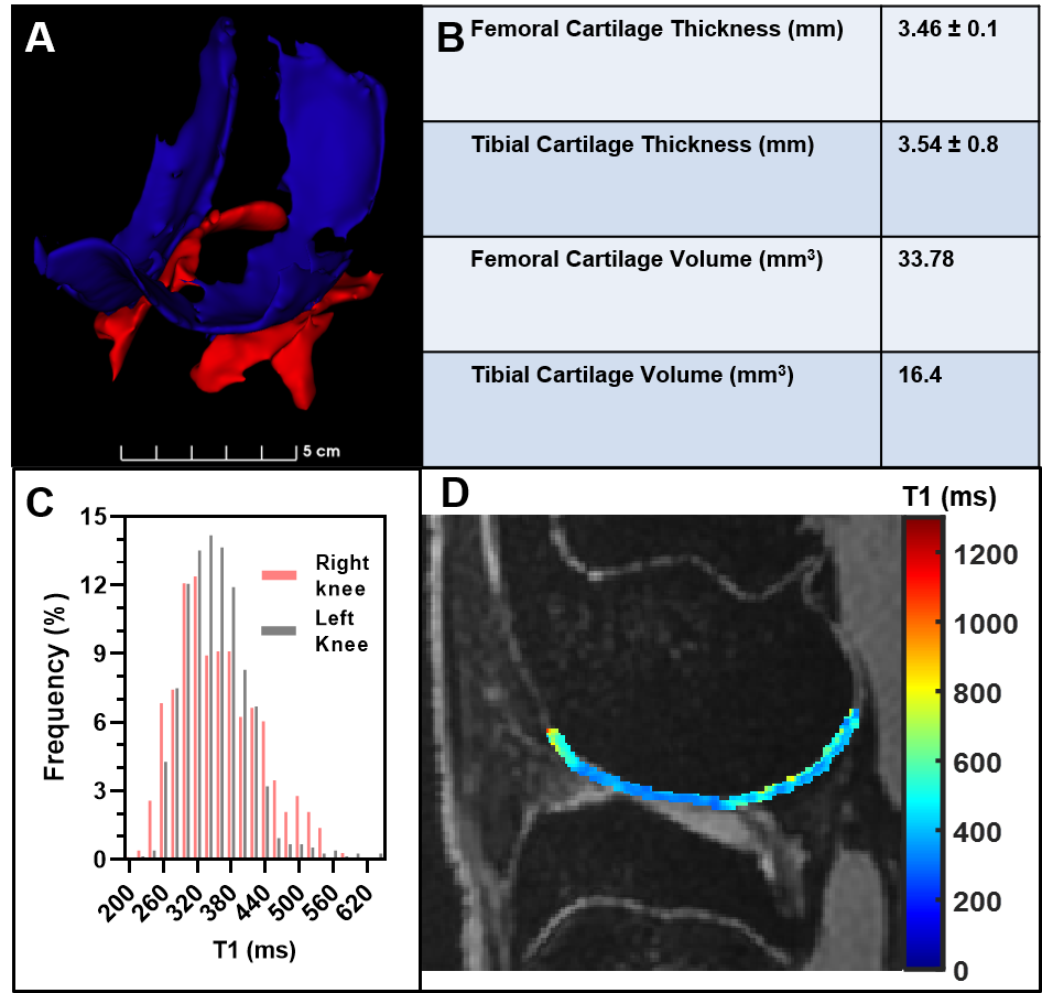
Cartilage
segmentation and quantification using the optimized protocols on both knees of
a healthy rabbit. (A) 3D map of the
femoral (blue) and tibial (red) cartilage, right knee. (B) Cartilage thickness
and volume quantification table, right knee.
(C) Histograms of T1 values from each knee joint showing the bilateral
similarity. (D) Relaxation map superimposed on one sagittal slice of DESS
image.
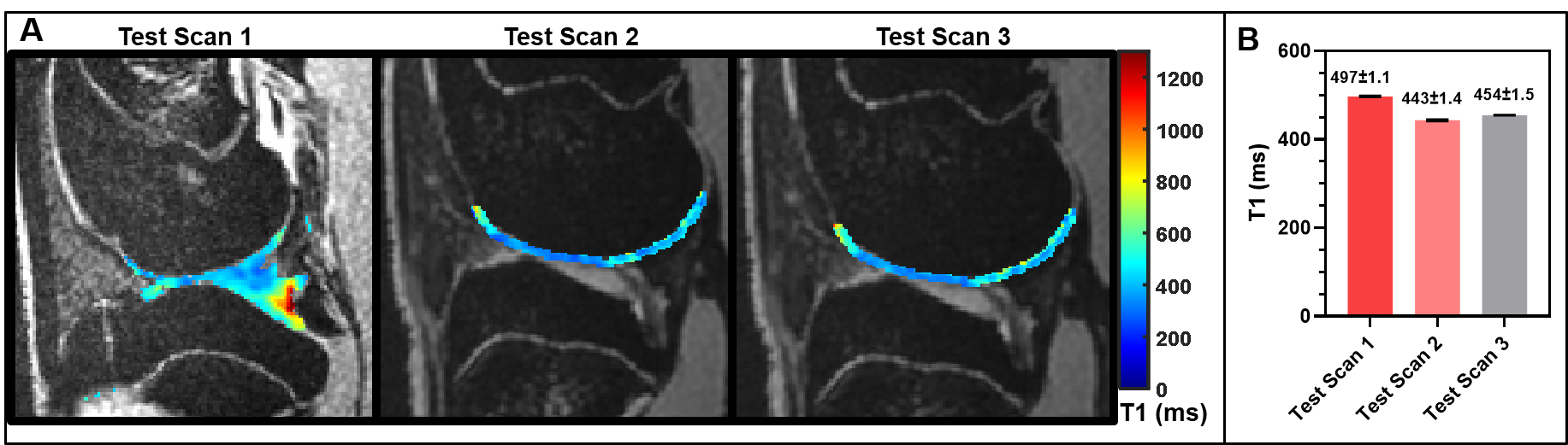
(A)
T1 maps on sagittal axis for three different MP2RAGE test scan conditions
(respectively: PAT factor 6, 3, 2, Echo Time: 3.85ms, 4.38ms, 4.38ms), superimposed
on the morphological DESS. ROIs are drawn in the cartilage 3-dimensionally
using a semi-automatic algorithm for segmentation on the DESS images. (B) Calculated
T1 values for three different MP2RAGE test scan conditions.