Zhenzhou Wu1, Stefan Sommer2,3, Xiaodong Zhong4, Kecheng Liu4, Jeehun Kim1, Jillian Beveridge1, Xiaoliang Zhang5, and Xiaojuan Li1
1Program of Advanced Musculoskeletal Imaging (PAMI), Cleveland Clinic, Cleveland, OH, United States, 2Siemens Healthcare, Zurich, Switzerland, 3Swiss Center for Musculoskeletal Imaging (SCMI), Balgrist Campus, Zurich, Switzerland, 4Siemens Medical Solutions USA, Inc., Malvern, PA, United States, 5Department of Biomedical Engineering, University at Buffalo, State University of New York, Buffalo, NY, United States
1Program of Advanced Musculoskeletal Imaging (PAMI), Cleveland Clinic, Cleveland, OH, United States, 2Siemens Healthcare, Zurich, Switzerland, 3Swiss Center for Musculoskeletal Imaging (SCMI), Balgrist Campus, Zurich, Switzerland, 4Siemens Medical Solutions USA, Inc., Malvern, PA, United States, 5Department of Biomedical Engineering, University at Buffalo, State University of New York, Buffalo, NY, United States
To evaluate
the repeatability of UTE T2*, and to investigate and compare the orientation
dependence of T2* mapping between UTE and regular gradient echo (GRE) imaging
sequences for whole knee imaging
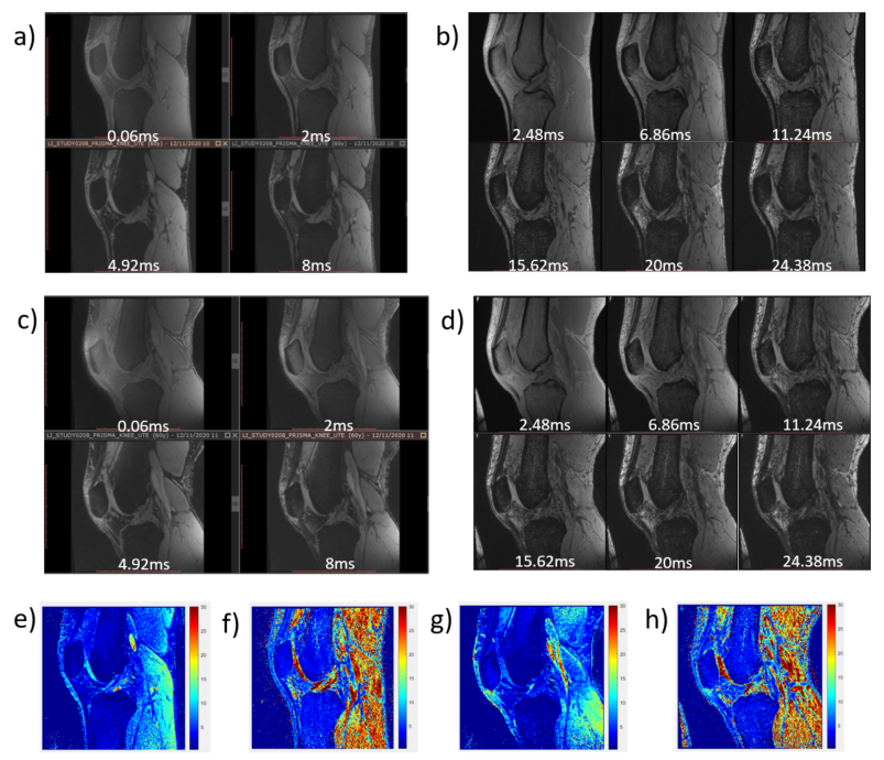
Figure 2. The 4 echoes of UTE and 6 echoes of GRE images with
different TE of extended (a and b) and flexed (c and d) knee. The
corresponding T2* mapping shown in e),
f), g), and h) respectively.
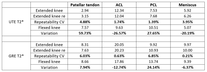
Table 3. In vivo repeatability and comparison of UTE and GRE
T2* relaxation time (in ms) of each compartment for both extended and flexed
knee joint.