Mei Wu1,2, Yajun Ma1, Guanyuan Ning2, Saeed Jerban1, Yanping Xue1, Zhao Wei1, Eric Y Chang1,3, and Jiang Du1
1Department of Radiology, University of California San Diego, San Diego, CA, United States, 2Department of Radiology, Guangzhou First People’s Hospital, School of Medicine, South China University of Technology, Guangzhou, China, 3Radiology Service, VA San Diego Healthcare System, San Diego, CA, United States
1Department of Radiology, University of California San Diego, San Diego, CA, United States, 2Department of Radiology, Guangzhou First People’s Hospital, School of Medicine, South China University of Technology, Guangzhou, China, 3Radiology Service, VA San Diego Healthcare System, San Diego, CA, United States
The 3D-UTE-Cones-AdiabT1ρ
sequence allow quantitative imaging of articular cartilage in the knee joint. The
3D-UTE-Cones-AdiabT1ρ values
are positively correlated with WORMS and
KL scores, and significantly different in different extent and depth lesions of
cartilage.
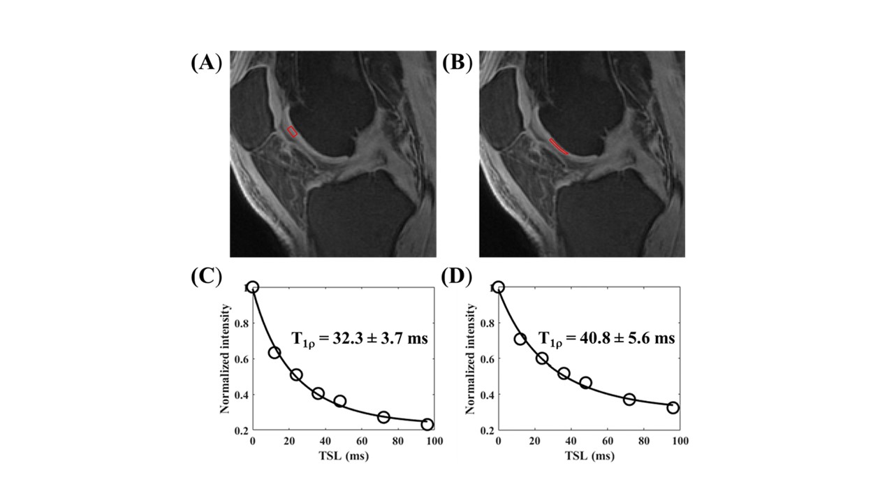
Figure 2. Excellent T1ρ modeling is
achieved for both normal cartilage (A, C) (T1ρ=32.3ms) and abnormal cartilage
(B, D) (WORMS=2, T1ρ=40.8ms).
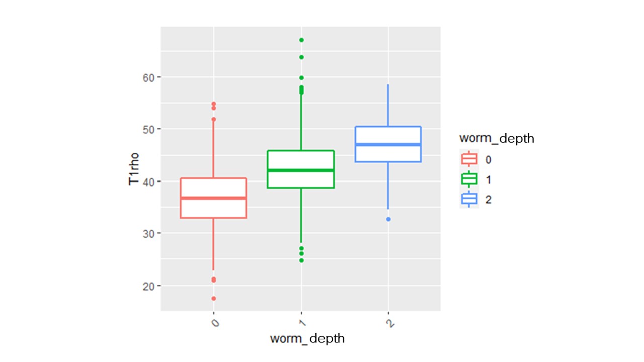
Figure 4. Boxplot
of UTE-Cones-AdiabT1ρ values in different WORMS depth groups