Takayuki Sakai1,2, Masami Yoneyama3, Atsuya Watanabe4,5, Daichi Murayama1, Shigehiro Ochi1, and Tosiaki Miyati6
1Radiology, Eastern Chiba Medical Center, Tonage, Japan, 2Division of Health Sciences, Graduate School of Medical Sciences, Kanazawa University, Kanazawa, Japan, 3Philips Japan, Tokyo, Japan, 4General Medical Services, Chiba University Graduate School of Medicine, Chiba, Japan, 5Orthopaedic Surgery, Eastern Chiba Medical Center, Chiba, Japan, 6Faculty of Health Sciences, Institute of Medical, Pharmaceutical and Health Sciences, Kanazawa University, Kanazawa, Japan
1Radiology, Eastern Chiba Medical Center, Tonage, Japan, 2Division of Health Sciences, Graduate School of Medical Sciences, Kanazawa University, Kanazawa, Japan, 3Philips Japan, Tokyo, Japan, 4General Medical Services, Chiba University Graduate School of Medicine, Chiba, Japan, 5Orthopaedic Surgery, Eastern Chiba Medical Center, Chiba, Japan, 6Faculty of Health Sciences, Institute of Medical, Pharmaceutical and Health Sciences, Kanazawa University, Kanazawa, Japan
SPLICE was combined with MultiVane (known as PROPELLER) which is to sample k-space in a rotating fashion using a set of radially directed blades. In this study, we evaluated the effect of accuracy and reproducibility of the quantitative values in lumbar nerve roots using MultiVane-SPLICE-DTI.
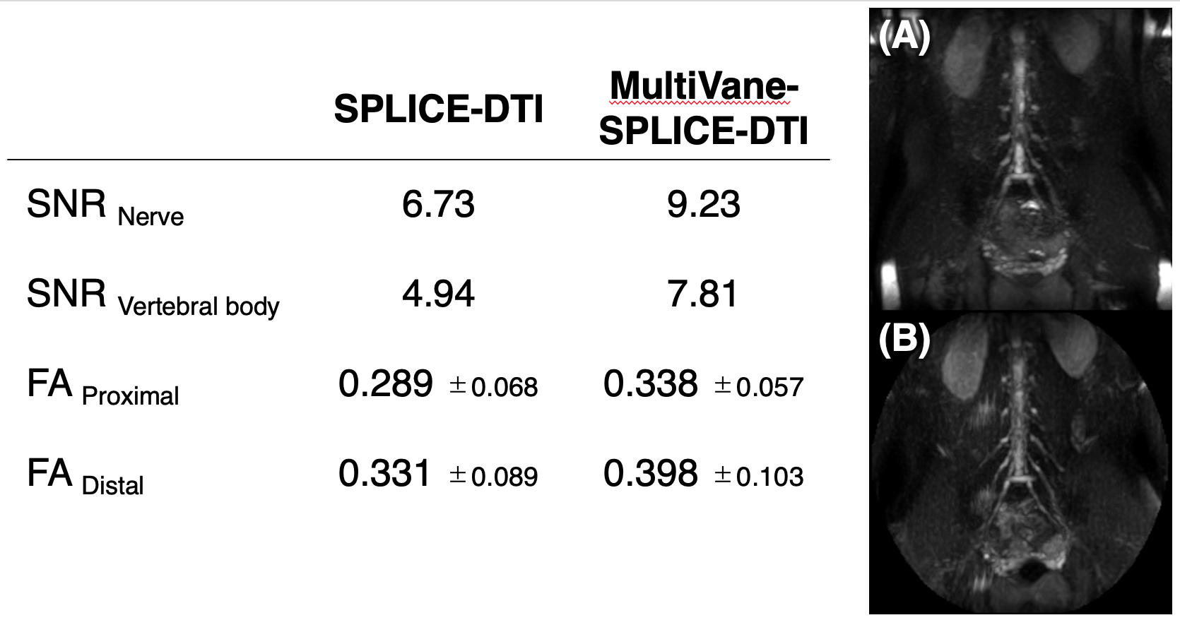
Fig.2 Comparison of SNR and FA values between SPLICE-DTI with MultiVane-SPLICE-DTI.
Partial Maximum Intensity Projection image (20mm thickness) shows on A (SPLICE-DTI) and B (MultiVane-SPLICE-DTI).
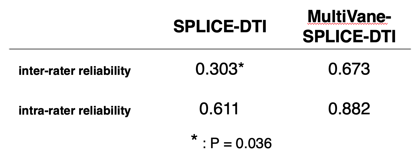
Fig.3 Comparison of ICC (inter-rater reliability and intra-rater reliability) between SPLICE-DTI with MultiVane-SPLICE-DTI in 3 healthy volunteers.