Shi Yin1, Dou Weiqiang2, and Ding Hongyuan1
1The First Affiliated Hospital of Nanjing Medical University, Nanjing, China, 2GE Healthcare, MR Research, Beijing, P.R. China, Beijing, China
1The First Affiliated Hospital of Nanjing Medical University, Nanjing, China, 2GE Healthcare, MR Research, Beijing, P.R. China, Beijing, China
Based on the characteristic pathological
change of nerve root edema in extraspinal sub-regions, conventional MR combined
with quantitative MR DTI/DTT has a good performance for visualizing and
quantitative diagnosis in lateral lumbar spinal canal stenosis.
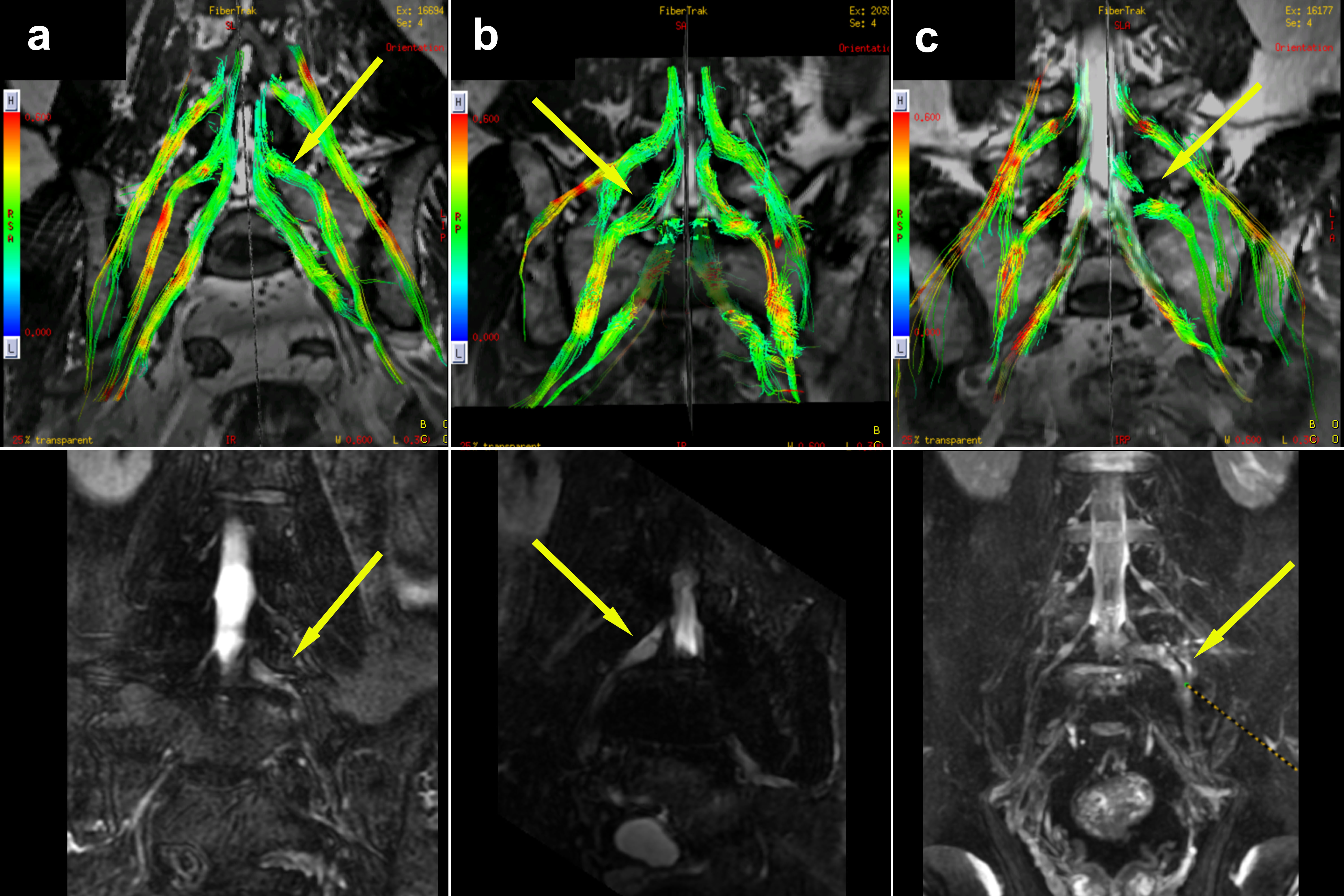
The DTT abnormalities of symptomatic nerves can
be classified into color hue reduction (a), sparse (b) and interruption
(c). The visual abnormalities of tractography were
found mainly in the extraforaminal (34 cases, 34.3%) and extraspinal (57 cases,
57.6%) region of compressed nerves.
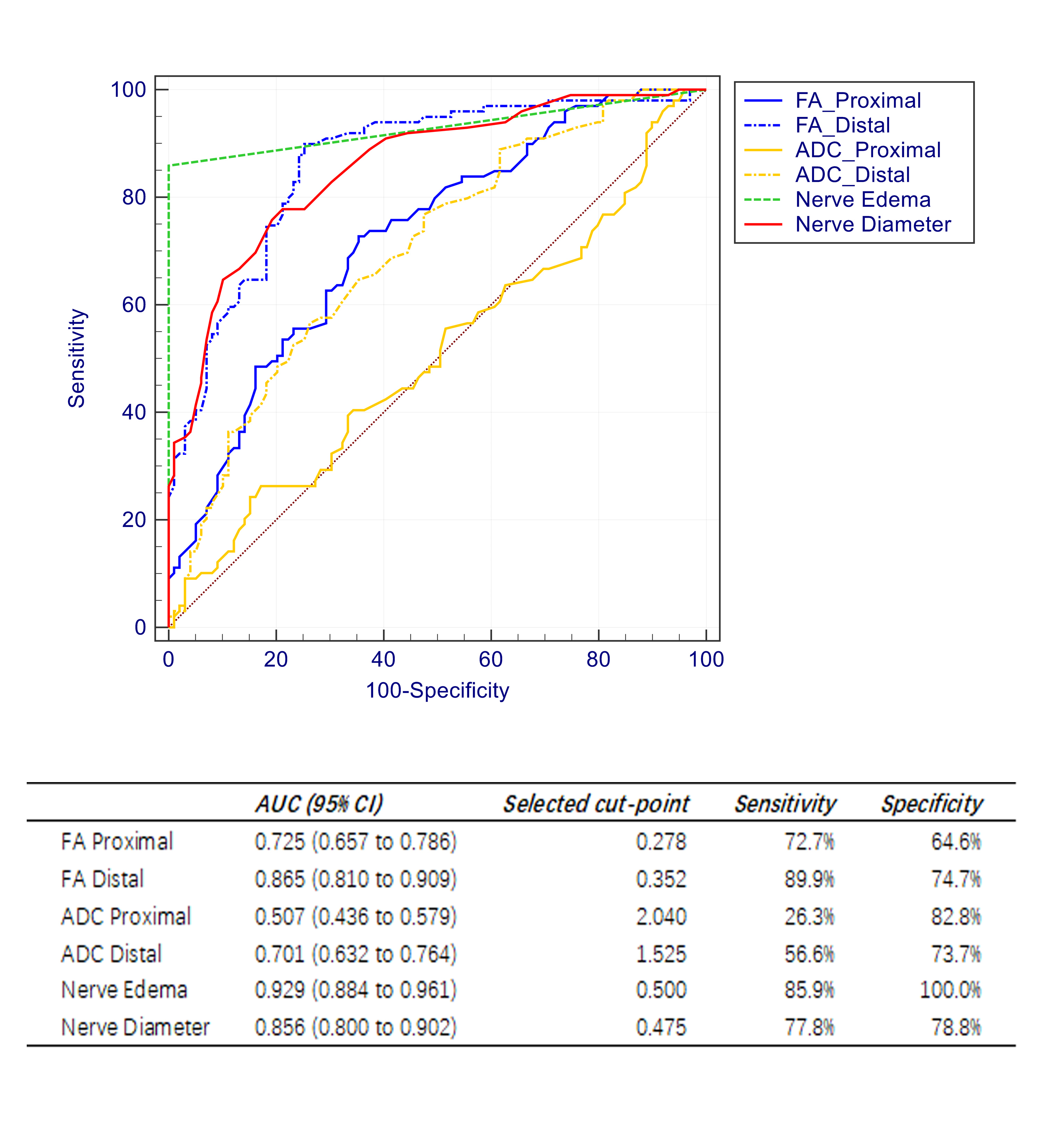
The ROC curve for nerve edema had the highest
AUC of 0.929 (95% CI 0.884 - 0.961); the cut point showed sensitivity and
specificity of 85.9% and 100%, respectively. In addition to this, the ROC curve
for distal FA and nerve diameter both had AUCs >0.80.