Jin Liu1, Yajun Ma2, Jianwei Liao1, Xiaojun Chen1, Wei Li1, Lin Yao1, Long Qian3, Jiang Du2, and Shaolin Li1
1Department of Radiology, The Fifth Affiliated Hospital of Sun Yat-Sen University, Zhuhai, China, 2Department of Radiology, University of California, San Diego, CA, United States, 3MR Research, GE Healthcare, Guangzhou, China
1Department of Radiology, The Fifth Affiliated Hospital of Sun Yat-Sen University, Zhuhai, China, 2Department of Radiology, University of California, San Diego, CA, United States, 3MR Research, GE Healthcare, Guangzhou, China
It
was concluded that IR-UTE was positively correlated to QCT and DXA, and was
negative correlated to FRAX scores. The 3D IR-UTE measures demonstrated good
performance in the differentiation of normal, osteopenia and osteoporosis.
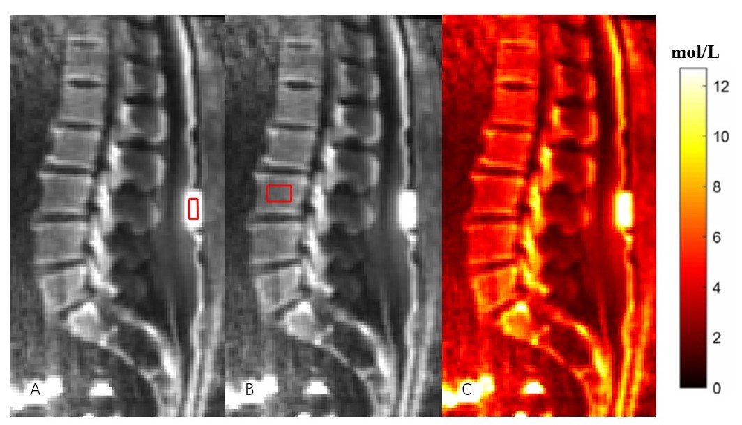
Figure 1: In vivo
qualitative and quantitative imaging of the lumbar of a 56-year-old female
volunteer using the 3D IR-UTE sequence. Figure
A: ROI
in the phantom;
Figure B: ROI in the lumbar vertebrae; Figure C: The proton density
map of the spine trabecular bone.
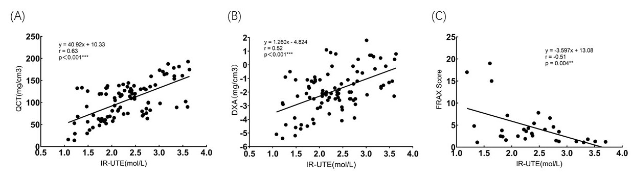
Figure 2: Linear regression in 30 subjects between (A)
IR-UTE measured PD and QCT, (B) IR-UTE measured PD and DXA, and (C) IR-UTE measured
PD and FRAX score.