Ek T Tan1, Erin C Argentieri1, Madeleine A Gao1, John Neri1, Garry E Gold2, Sharmila Majumdar3, Hollis G Potter1, and Matthew F Koff1
1Radiology and Imaging, Hospital for Special Surgery, New York, NY, United States, 2Stanford University, Stanford, CA, United States, 3University of California San Francisco, San Francisco, CA, United States
1Radiology and Imaging, Hospital for Special Surgery, New York, NY, United States, 2Stanford University, Stanford, CA, United States, 3University of California San Francisco, San Francisco, CA, United States
T2* mapping with UTE may provide increased sensitivity to subclinical changes in the collagen matrix of menisci. Denoising of high-resolution, low-SNR images can reduce variance of T2* maps, leading to improved qMRI.
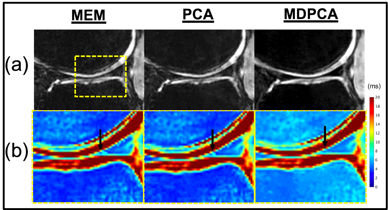
Fig.1. (a) UTE (TE=30ms) images, showing reduced noise with PCA and MDPCA, and (b) improved fitting error with MDPCA in the T2* maps of cropped region corresponding to dashed box region (arrow – posterior horn of medial meniscus).
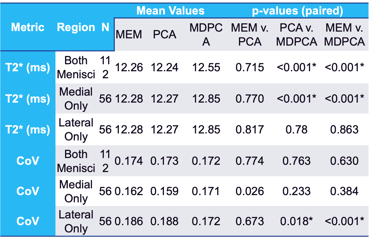
Table 1. Paired
Comparisons of Mean T2* and Coefficient of Variation (CoV) for MEM, PCA-denoising
and MD+PCA-denoising Algorithms. Note: * = p < 0.05.