Varvara Choida1,2, Timothy J.P. Bray1,3, Alan Bainbridge4, Debajit Sen2,5, Corinne Fisher2,5, Maria Leandro2,5, Coziana Ciurtin2,5, and Margaret Hall-Craggs1,3
1Centre for Medical Imaging, University College London, London, United Kingdom, 2Centre for Adolescent Rheumatology Versus Arthritis at UCL UCLH and GOSH, University College London, London, United Kingdom, 3Radiology, University College London Hospital, London, United Kingdom, 4Medical Physics, University College London Hospitals, London, United Kingdom, 5Adolescent and young adult Rheumatology, University College London Hospital, London, United Kingdom
1Centre for Medical Imaging, University College London, London, United Kingdom, 2Centre for Adolescent Rheumatology Versus Arthritis at UCL UCLH and GOSH, University College London, London, United Kingdom, 3Radiology, University College London Hospital, London, United Kingdom, 4Medical Physics, University College London Hospitals, London, United Kingdom, 5Adolescent and young adult Rheumatology, University College London Hospital, London, United Kingdom
We developed a whole-body MRI protocol, consisting of post-contrast mDixon images, which revealed a large burden of clinically undetected synovitis in 32 adolescent patients with clinically active and inactive juvenile idiopathic arthritis.
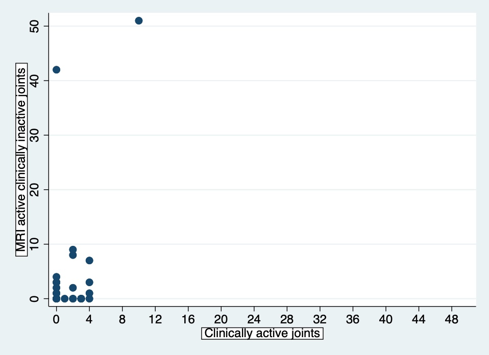
Figure 4. Synovitis detected by MRI in clinically inactive joints versus clinical synovitis. Values represent number of joints per patient. In many patients, there are additional joints with synovitis on MRI that were clinically unsuspected.
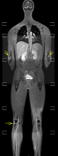
Figure 1. Bilateral elbow and right knee synovitis on post-contrast, water-only Dixon MR image (whole-body view).