Jacob Thoenen1, James W. MacKay2,3, Kathryn J. Stevens1, Tom D. Turmezei4, Akshay Chaudhari1, Lauren E. Watkins1, Emily J. McWalter5, Brian A. Hargreaves1, Garry E. Gold1, and Feliks Kogan1
1Department of Radiology, Stanford University, Stanford, CA, United States, 2Department of Radiology, University of Cambridge, Cambridge, United Kingdom, 3Norwich Medical School, University of East Anglia, Norwich, United Kingdom, 4Department of Radiology, Norfolk and Norwich University Hospital, Norwich, United Kingdom, 5Department of Mechanical Engineering, University of Saskatchewan, Saskatoon, SK, Canada
1Department of Radiology, Stanford University, Stanford, CA, United States, 2Department of Radiology, University of Cambridge, Cambridge, United Kingdom, 3Norwich Medical School, University of East Anglia, Norwich, United Kingdom, 4Department of Radiology, Norfolk and Norwich University Hospital, Norwich, United Kingdom, 5Department of Mechanical Engineering, University of Saskatchewan, Saskatoon, SK, Canada
The quantitative double echo in steady state (qDESS) sequence shows good agreement to contrast-enhanced MRI for characterization of the severity of synovitis.
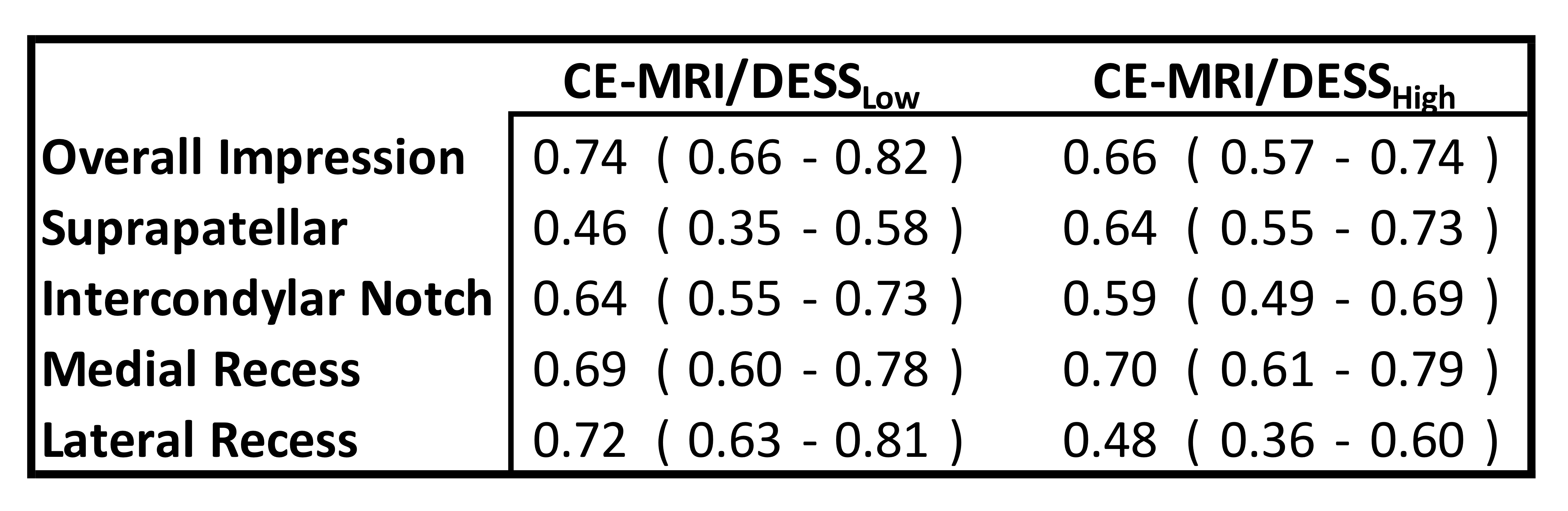
Table 2: Agreement between qDESS and CE-MRI using Gwet's AC2 (95% LOA) for readers overall impression of synovitis and regional gradings
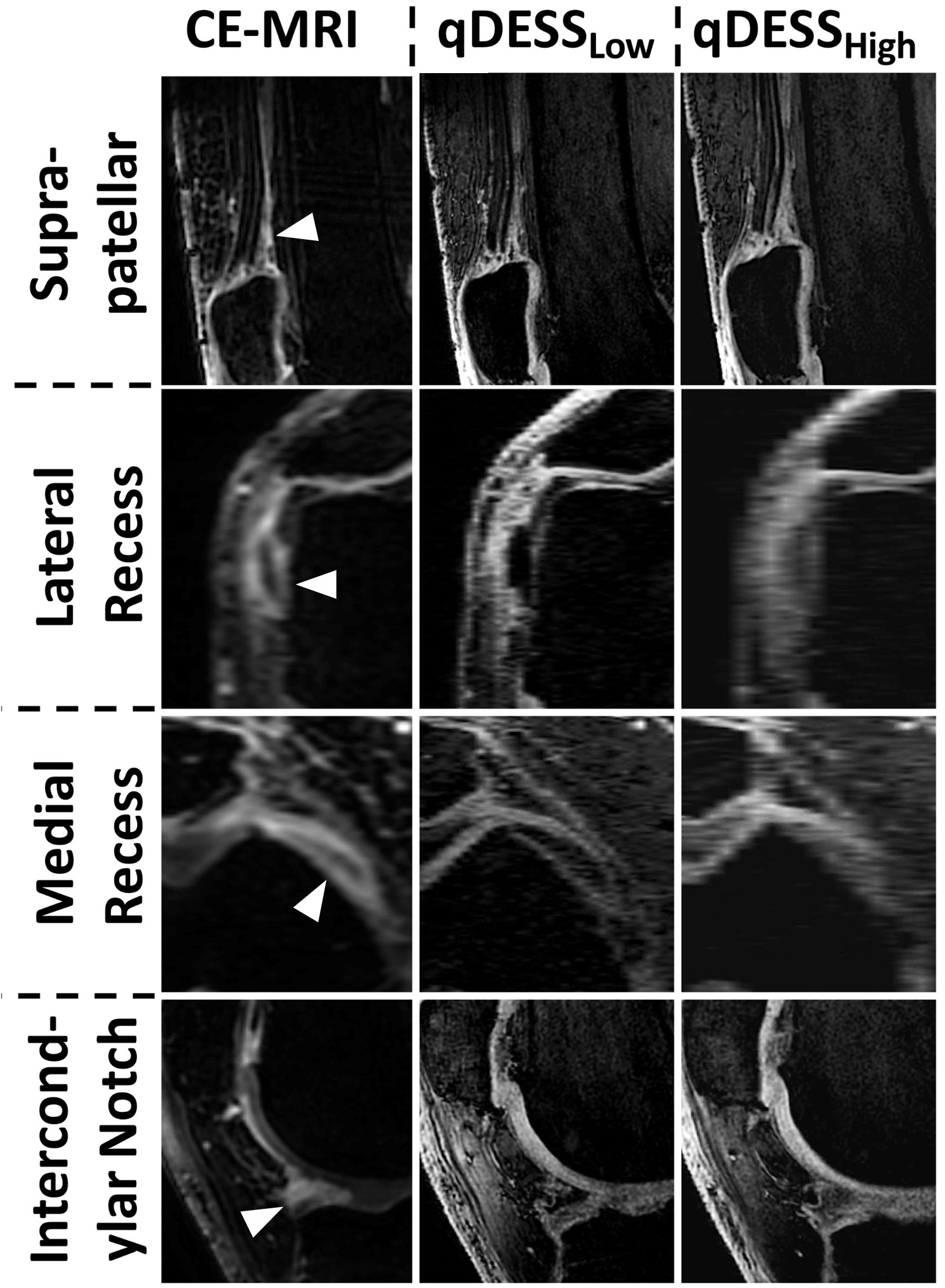
Figure 1: Example cases where radiologists tended to agree on severity of synovitis on CE-MRI, qDESSLow, and qDESSHigh. In the sagittal MR images of the suprapatellar pouch, mild uniform synovial thickening is demonstrated without obvious nodularity (grade 1). In the axial MR images of the medial and lateral recess, synovial thickening is more conspicuous with the suggestion of nodularity in the lateral recess images (grade 2). In the sagittal intercondylar notch images, there is nodular synovial enhancement anterior to the anterior horn of the lateral meniscus (grade 2)