Erica Silvestri1,2,3, Marco Castellaro1,4, Alessandra Zorz5, Andrea Bettinelli 1,5, Cristina Campi6, Marta Paiusco5, Diego Cecchin2,7, and Alessandra Bertoldo1,2
1Department of Information Engineering, University of Padova, Padova, Italy, 2Padova Neuroscience Center, University of Padova, Padova, Italy, 3Department of Neuroscience, University of Padova, Padova, Italy, 4Department of Neurosciences, Biomedicine and Movement Sciences, University of Verona, Verona, Italy, 5Department of Medical Physics, Veneto Institute of Oncology IOV - IRCCS, Padova, Italy, 6Department of Math, University of Padova, Padova, Italy, 7Department of Medicine, Unit of Nuclear Medicine, University of Padova, Padova, Italy
1Department of Information Engineering, University of Padova, Padova, Italy, 2Padova Neuroscience Center, University of Padova, Padova, Italy, 3Department of Neuroscience, University of Padova, Padova, Italy, 4Department of Neurosciences, Biomedicine and Movement Sciences, University of Verona, Verona, Italy, 5Department of Medical Physics, Veneto Institute of Oncology IOV - IRCCS, Padova, Italy, 6Department of Math, University of Padova, Padova, Italy, 7Department of Medicine, Unit of Nuclear Medicine, University of Padova, Padova, Italy
HD-EEG net attenuates PET signal in NAC images in both phantom and human brain, respectively of 3.01% (±3.69%) and of 0.53% (±5.89%). AC images show an opposite behaviour: attenuation correction factor tends to be overestimated in CT- and UTE-derived UMAP obtained with HD-EEG net.
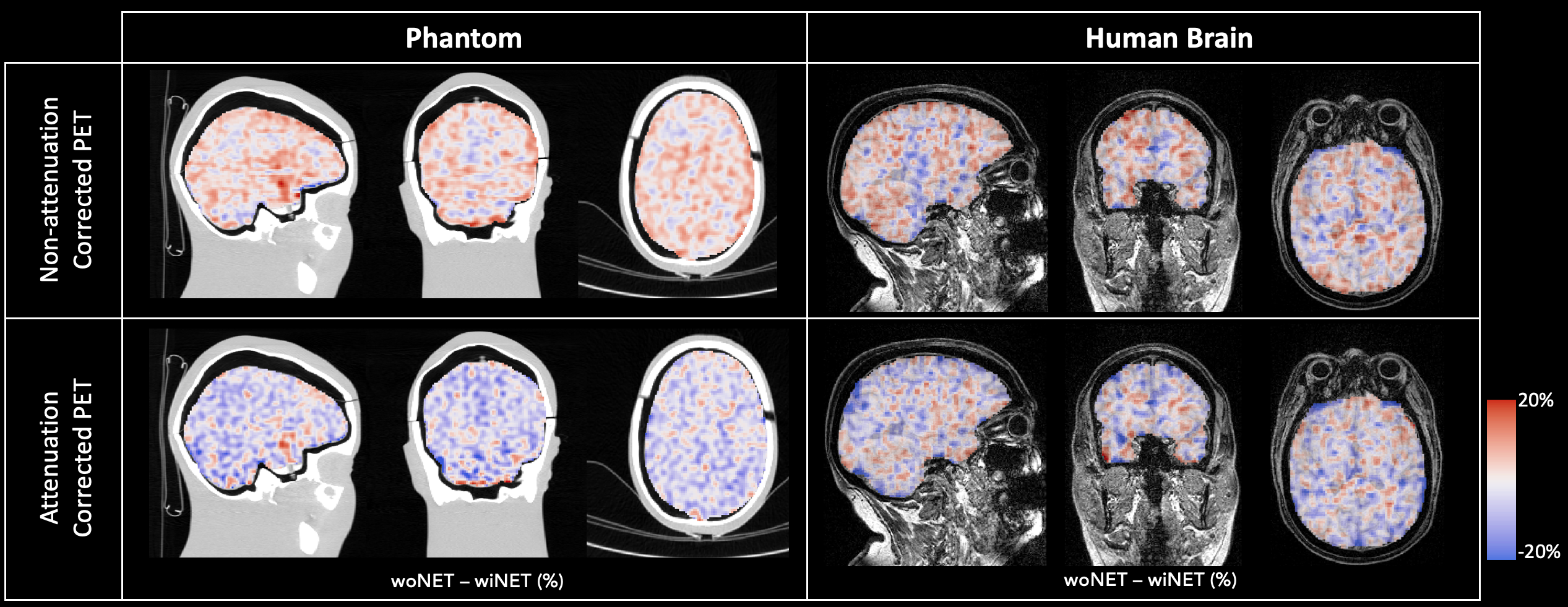
Figure 3: Spatial pattern of relative % differences between PET images acquired with and without HD-EEG net (blue-red scale) over-imposed on phantom CT image or human brain T1w MPRAGE image (grey scale).
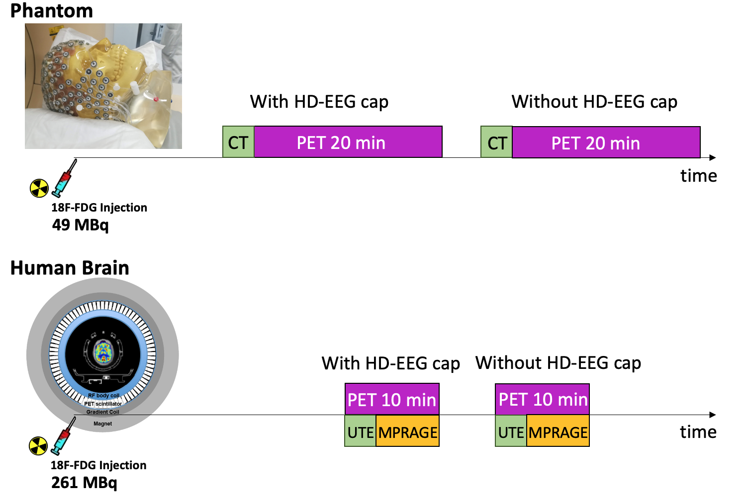
Figure 1: Schematic representation of the two acquisition protocols.