Rakshit Dadarwal1,2, Fabien Balezeau3, Marcus Haag4, Michael C. Schmid3,4, Susann Boretius1,2, and Michael Oritz-Rios1,3
1Functional Imaging Laboratory, German Primate Center, Göttingen, Germany, 2Georg August Universität Göttingen, Göttingen, Germany, 3Biosciences Institute, Newcastle University, Newcastle upon Tyne, United Kingdom, 4Faculty of Science and Medicine, University of Fribourg, Fribourg, Switzerland
1Functional Imaging Laboratory, German Primate Center, Göttingen, Germany, 2Georg August Universität Göttingen, Göttingen, Germany, 3Biosciences Institute, Newcastle University, Newcastle upon Tyne, United Kingdom, 4Faculty of Science and Medicine, University of Fribourg, Fribourg, Switzerland
In this work, we demonstrate the feasibility and robustness of multi-contrast MRI data acquisition across sessions and subjects in awake macaque monkeys.
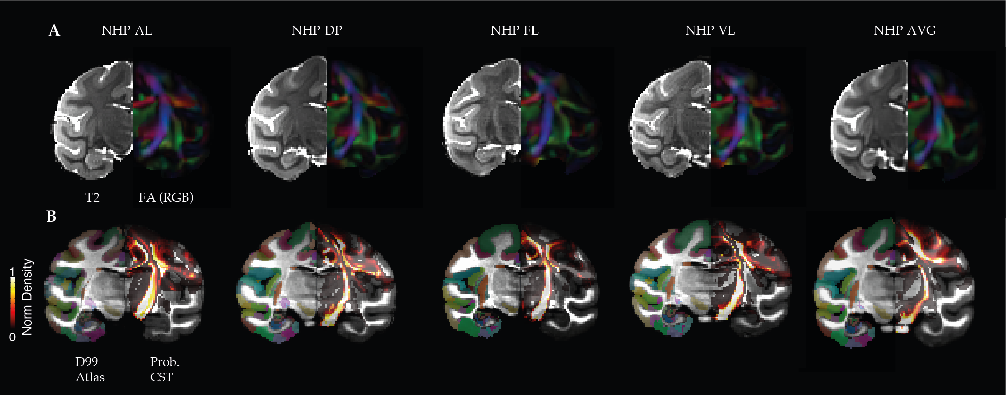
Figure 1. Example coronal slices for each NHPs and the subject average template. A. Left and right pairs slices showing the within-subject average for each type of dataset, T2w and color-coded fractional anisotropy (FA-RGB). B. Left and right pairs slices showing the individually warped D99 atlas (left) and the resulting probabilistic map showing the normalized fiber density of the cortico-spinal tract (CST).
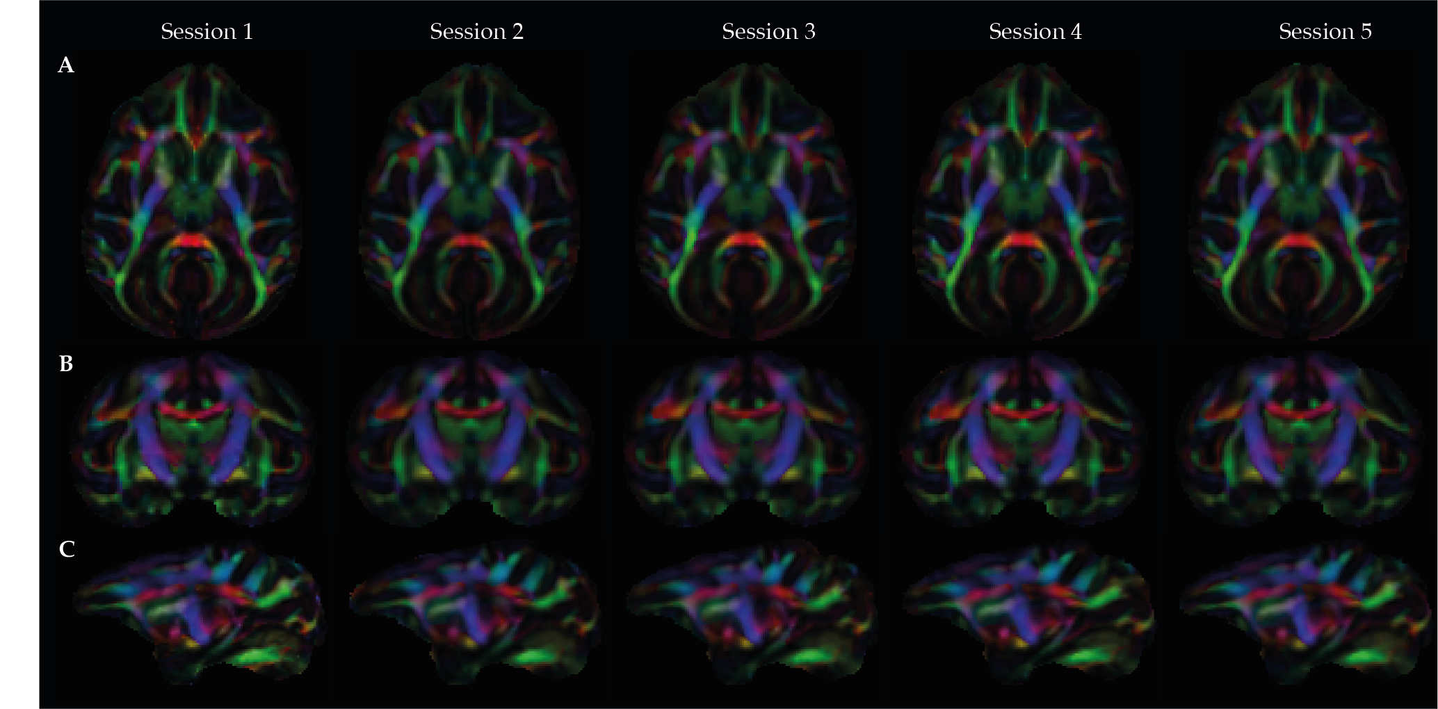
Figure 2. Intra-subject reproducibility analysis for the color-coded FA maps in axial (A), coronal (B), and sagittal (C) planes. The same subject (AL) was scanned five times (Session 1 to Session 5) using the same acquisition protocol. The color-coding shows white matter fiber tracts in red (left to right), green (anterior to posterior), and blue (dorsal to ventral) orientations.