Suk-tak Chan1, Nathaniel Mercaldo2, Kenneth K. Kwong1, Steven M. Hersch3, and Herminia D. Rosas3
1Athinoula A. Martinos Center for Biomedical Imaging, Department of Radiology, Massachusetts General Hospital, Charlestown, MA, United States, 2Department of Radiology, Massachusetts General Hospital, Boston, MA, United States, 3Department of Neurology, Massachusetts General Hospital, Boston, MA, United States
1Athinoula A. Martinos Center for Biomedical Imaging, Department of Radiology, Massachusetts General Hospital, Charlestown, MA, United States, 2Department of Radiology, Massachusetts General Hospital, Boston, MA, United States, 3Department of Neurology, Massachusetts General Hospital, Boston, MA, United States
Alterations in
cerebrovascular function was found in HD and the dominance of such alterations in
white matter further suggests the signs of small vessel
disease. The impaired cerebrovascular
reactivity may be an important, not as yet considered, contributor to early
neuropathology in HD.
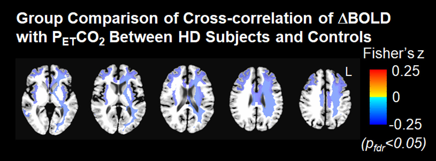
Figure 1. Group comparison of cross-correlation of ΔBOLD
with PETCO2 between HD subjects and healthy controls
after adjusting for age, corrected at pfdr<0.05. Cold colors represent weaker
cross-correlation in HD subjects when compared with controls.
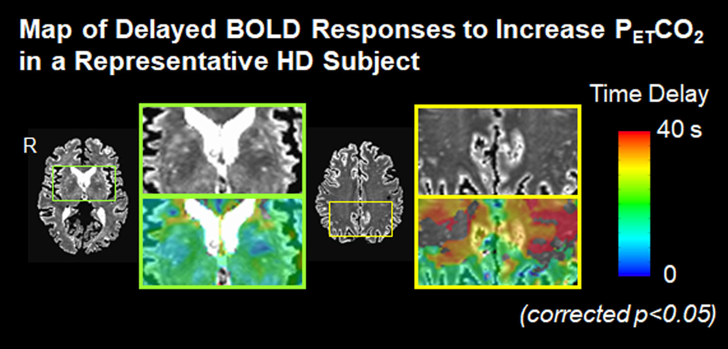
Figure 2. Map of delayed BOLD responses to increased PETCO2
in a representative HD subject. The
magnitude of time delay in BOLD responses relative to PETCO2
increases from cold colors to warm colors.