Andrew Crofts1, Jessica J Steventon2, Joseph R Whittaker3, Marcello Venzi1, Hannah L Chandler4, Mahfoudha Al Shezawi5, Eric J Stohr6, Chris Pugh6, Barry McDonnell6, and Kevin Murphy1
1Cardiff University Brain Research Imaging Centre (CUBRIC), School of Physics and Astronomy, Cardiff University, Cardiff, United Kingdom, 2CUBRIC, School of Medicine, Cardiff University, Cardiff, United Kingdom, 3Max Planck Institute for Human Cognitive and Brain Sciences, Leipzig, Germany, 4CUBRIC, School of Psychology, Cardiff University, Cardiff, United Kingdom, 5Cardiff Metropolitan University, Cardiff, United Arab Emirates, 6Cardiff Metropolitan University, Cardiff, United Kingdom
1Cardiff University Brain Research Imaging Centre (CUBRIC), School of Physics and Astronomy, Cardiff University, Cardiff, United Kingdom, 2CUBRIC, School of Medicine, Cardiff University, Cardiff, United Kingdom, 3Max Planck Institute for Human Cognitive and Brain Sciences, Leipzig, Germany, 4CUBRIC, School of Psychology, Cardiff University, Cardiff, United Kingdom, 5Cardiff Metropolitan University, Cardiff, United Arab Emirates, 6Cardiff Metropolitan University, Cardiff, United Kingdom
Central
pulse pressure and its influence on carotid artery morphology better predict
white matter hyperintensity volume than both brachial blood pressure and pulse
wave velocity, a measure of arterial stiffness.
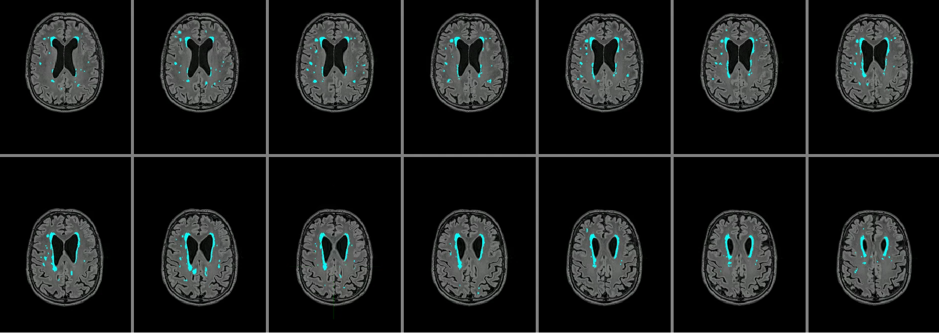
Figure 3: Example image showing large white matter hyperintensities
(blue) overlaid on the subject’s FLAIR image. The total white matter hyperintensities volume for
this subject was 13406 voxels.
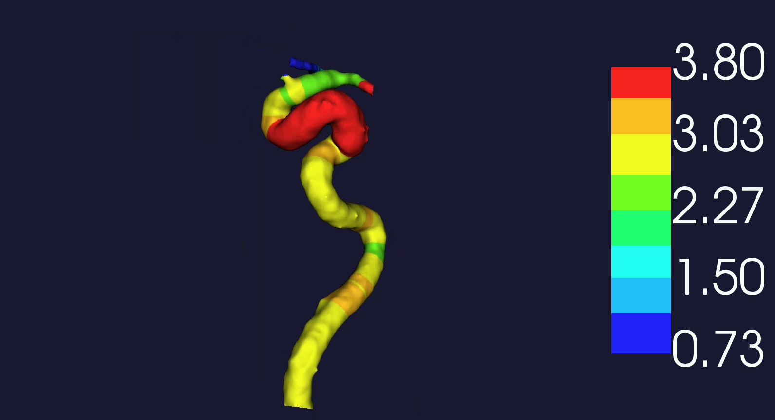
Figure 4: Example
image showing the left carotid artery from the same subject as Figure 3. The
colour map shows the arterial radius (average across the vessel = 2.9470). The
tortuosity for this vessel was 0.8773.