Shoko Hara1,2, Masaaki Hori2,3, Yoji Tanaka1, Taketoshi Maehara1, Shigeki Aoki2, and Tadashi Nariai1
1Neurosurgery, Tokyo Medical and Dental University, Tokyo, Japan, 2Radiology, Juntendo University, Tokyo, Japan, 3Radiology, Toho University Omori Medical Center, Tokyo, Japan
1Neurosurgery, Tokyo Medical and Dental University, Tokyo, Japan, 2Radiology, Juntendo University, Tokyo, Japan, 3Radiology, Toho University Omori Medical Center, Tokyo, Japan
We analyzed 13 patients with moyamoya disease before and after the revascularization surgery by multishell diffusion MRI. By NODDI and free-water imaging analysis, increased extracellular fluid showed significant decrease after the surgery.
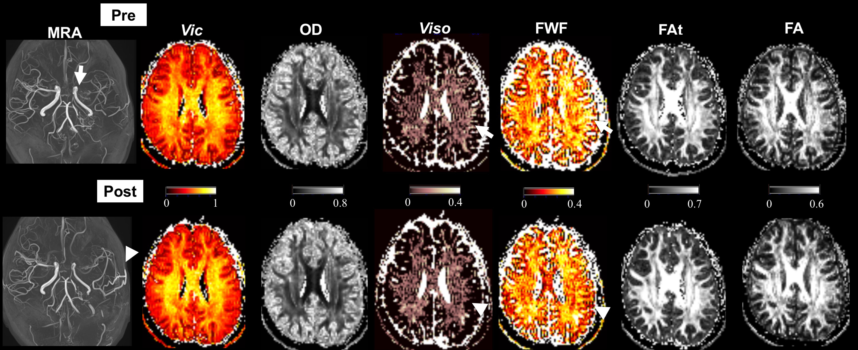
Figure 1. Parametrical maps of the representative case. A 19 year-old female who presented with transcient ischemic attacks of the right side received revascularization surgery (indirect bypass surgery) to the left hemisphere. Preoperative isotropic volume fraction (Viso) of NODDI and freewater fraction (FWF) of regularized bi-tensor model suggests increased water component in the left hemisphere (arrows) compared to the contralateral hemisphere. The increased water seems to decrease after the surgery (arrows).
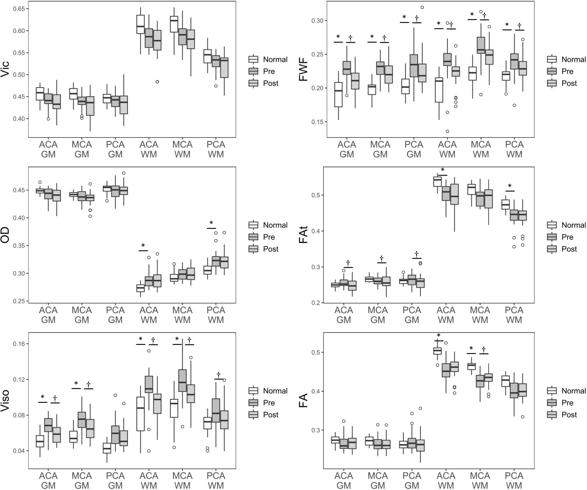
Parametrical values of normal controls (Normal), preoperative (Pre) and postoperative (Post) patients in six regions. Compared to normal controls, preoperative regions showed higher Viso and FWF in most regions, and significantly decreased after the surgery. In the white matter (WM) of the ACA areas, OD is higher, and FAt and FA are lower compared than normal controls. FA in the WM of MCA showed significant increase after the surgery. GM, gray matter. *P<.05 (unpaired T test) and †P<.05 (paired T test) after Bonferroni correction for six parameters.