Alexandra Ereni King1, Paul D Teal2, Yu-Chieh Tzeng3, Annabel Jain Sorby-Adams4, Isabella Megan Bilecki4, Renée Jade Turner4, and Sergei Obruchkov5
1School of Chemical and Physical Sciences, Victoria University of Wellington, Wellington, New Zealand, 2School of Engineering and Computer Science, Victoria University of Wellington, Wellington, New Zealand, 3Centre for Translational Physiology, University of Otago, Wellington, New Zealand, 4Discipline of Anatomy and Pathology, University of Adelaide, Adelaide, Australia, 5Robinson Research Institute, Victoria University of Wellington, Wellington, New Zealand
1School of Chemical and Physical Sciences, Victoria University of Wellington, Wellington, New Zealand, 2School of Engineering and Computer Science, Victoria University of Wellington, Wellington, New Zealand, 3Centre for Translational Physiology, University of Otago, Wellington, New Zealand, 4Discipline of Anatomy and Pathology, University of Adelaide, Adelaide, Australia, 5Robinson Research Institute, Victoria University of Wellington, Wellington, New Zealand
Quantitative perfusion parameters (e.g. volume transfer coefficient, interstitial and plasma volume fractions) in a novel ovine model of stroke are calculated from DCE-MRI images of the healthy animal, at 24 hours, 3 days, 6 days and 28 days post stroke.
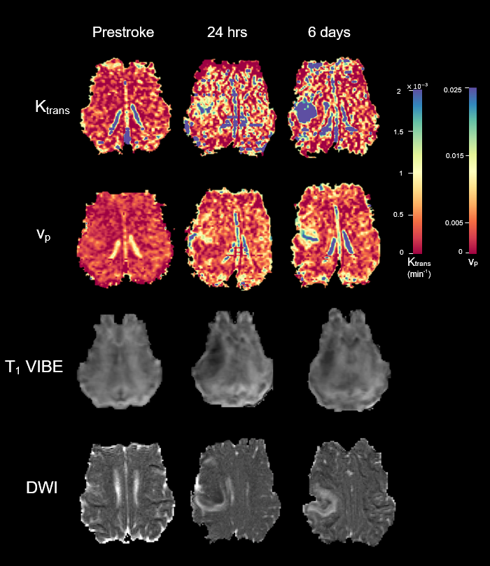
Perfusion map of Ktrans [min-1] (top row) and vp [arb.] (second row), T1 weighted images (third row), and diffusion weighted images (bottom row) of a single animal from before stroke, 24 hours post stroke, and 6 days post stroke.