Maoxue Wang1, Yongbo Yang1, Fei Zhou1, Ming Li1, Jilei Zhang2, and Bing Zhang1,3
1The Affiliated Drum Tower Hospital of Nanjing University Medical School, Nanjing, China, 2Philips Healthcare, Shanghai, China, 3Institute of Brain Science, Nanjing University Nanjing, Nanjing, China
1The Affiliated Drum Tower Hospital of Nanjing University Medical School, Nanjing, China, 2Philips Healthcare, Shanghai, China, 3Institute of Brain Science, Nanjing University Nanjing, Nanjing, China
The
mean value of CBF in cerebral hemispheres with lesions was lower than that on
the contralateral side in ASL with PLDs of 1.5 s and 2.5 s, and it was correlated
with the occurrence of cerebrovascular events.
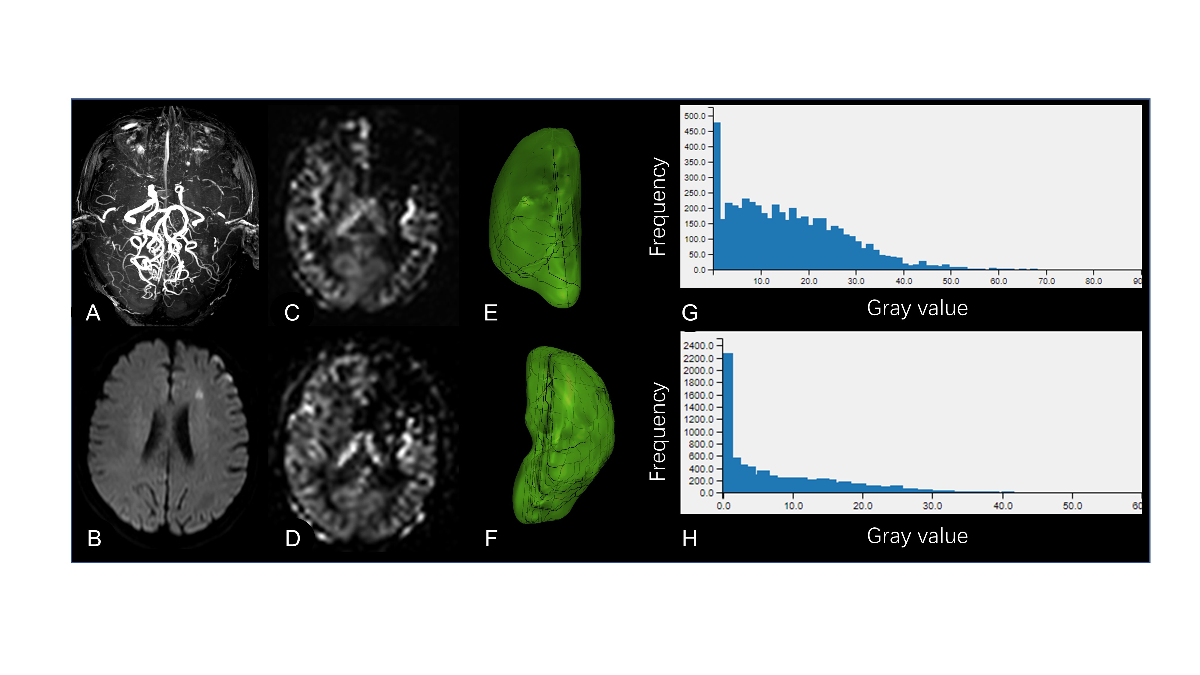
Figure 1. A 61-year-old patient with bilateral MMA. (A)
Occlusion of bilateral ICA, ACA, and MCA was observed on 3D time of flight MRA
images. (B) He had a punctate acute cerebral infarction in the left frontal
lobe. The
perfusion of the left frontal lobe decreased, and the arterial transit artifact
was grade 2 on CBF images when PLD were 1.5 s and 2.5 s (C and D). (E and F) Three-dimensional
images of bilateral cerebral hemispheres. The gray value of the left cerebral
hemisphere was less than that of the contralateral on the gray distribution
histogram of bilateral cerebral hemispheres.
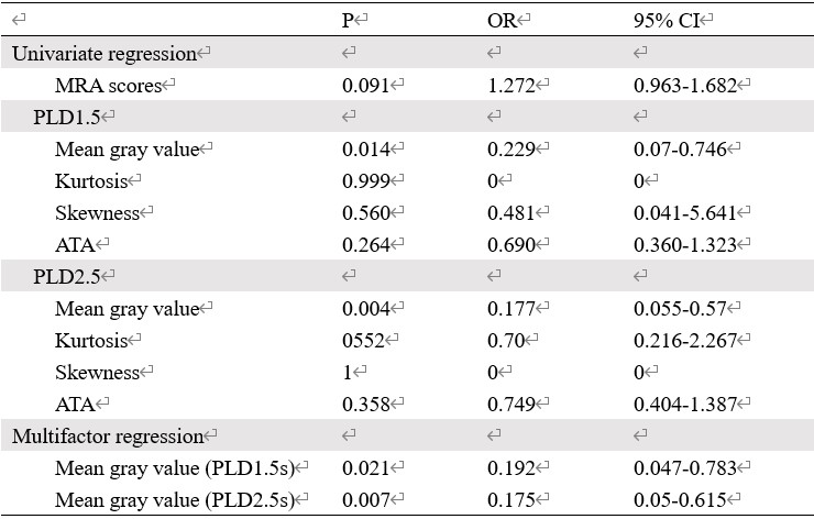
Table 2. Correlation between the degree of
intracranial artery, cerebral perfusion, and occurrence of cerebrovascular
events in patients with MMA.